
Axon Collection
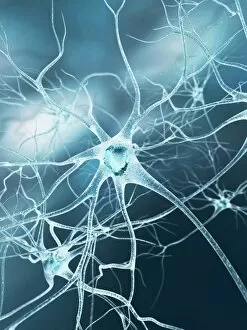
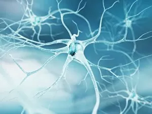
The intricate world of the axon, a vital component of nerve cells, comes to life through advanced imaging techniques


All Professionally Made to Order for Quick Shipping
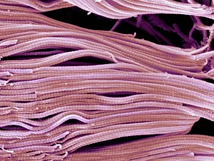
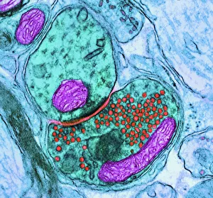
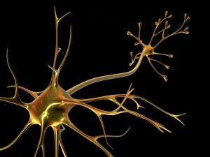
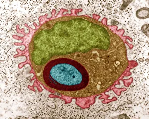
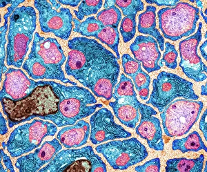
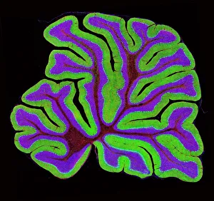
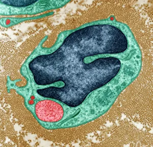
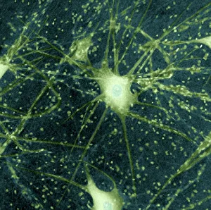
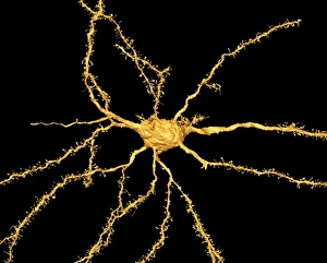
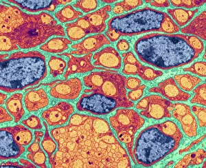
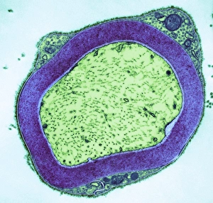
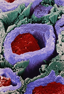
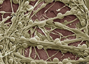
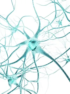
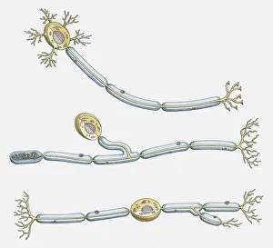
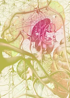
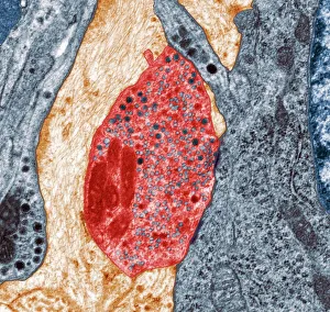
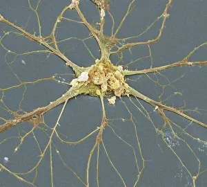
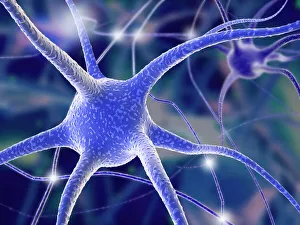
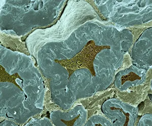
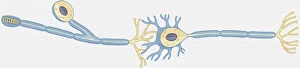
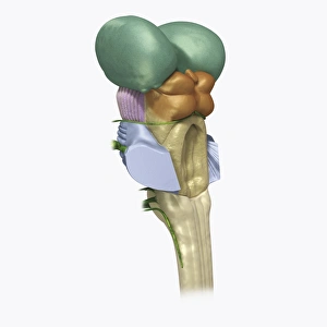
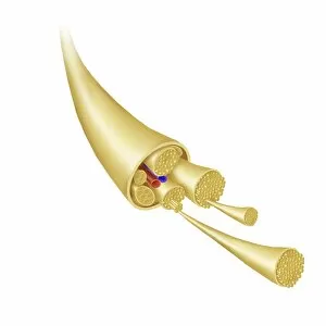
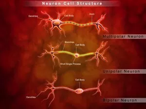
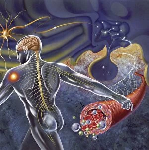
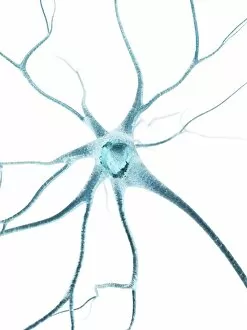
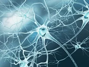
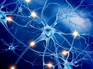
The intricate world of the axon, a vital component of nerve cells, comes to life through advanced imaging techniques. In the synapse nerve junction, where communication between neurons occurs, transmission electron microscopy (TEM) reveals a mesmerizing web of connections. Collagen fibers intertwine with delicate axons in a scanning electron micrograph (SEM), highlighting their structural support. Regeneration is key for damaged nerves, and TEM captures the awe-inspiring process. A regenerating nerve cell emerges from its cocoon-like state, showcasing its resilience and potential for healing. Myelination of nerve fibers is another fascinating phenomenon observed under TEM; this protective sheath enhances signal conduction efficiency. Zooming out to explore larger structures within our brain, light micrographs unveil the complexity of the cerebellum's architecture. Its convoluted layers resemble an abstract masterpiece waiting to be deciphered. Motor neurons also take center stage in these images; their distinctive shapes hint at their crucial role in controlling movement. Returning to TEM imagery once more, we witness yet another regenerating nerve cell undergoing transformation—a testament to nature's remarkable ability to restore itself. Synapse nerve junctions are further examined using SEM, revealing an intricately woven network that facilitates rapid information transfer between neurons. Delving deeper into the microscopic realm with SEM unveils stunning images of individual nerve cells—each one unique and essential for proper brain function. The myelination process continues captivating us as TEM showcases its intricate details once again. To better understand neuron diversity, an illustration presents multipolar, unipolar, and bipolar neurons side by side—an educational glimpse into their distinct characteristics and functions within our nervous system.