
Axoneme Collection
The axoneme is a crucial component of human spermatids, responsible for the formation of sperm tails


All Professionally Made to Order for Quick Shipping
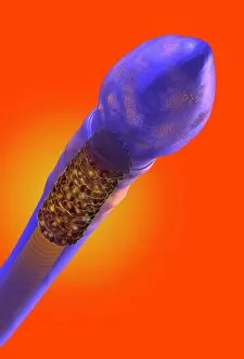
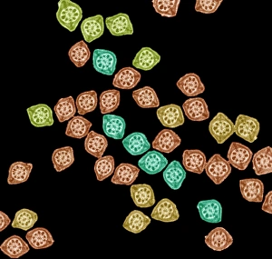
The axoneme is a crucial component of human spermatids, responsible for the formation of sperm tails. Through Transmission Electron Microscopy (TEM), scientists have been able to capture detailed images of this intricate structure. In artwork C018 / 6996, we can see a beautifully illustrated sperm cell with its axoneme highlighted. This microscopic powerhouse propels the sperm forward, aiding in fertilization. TEM images such as C014 / 1463, C014 / 1465, and C014 / 1464 provide us with an up-close look at the complexity of sperm tails. These slender structures consist of microtubules arranged in a specific pattern that allows for efficient movement. Artwork C013 / 4648 showcases the anatomy of a sperm cell, highlighting not only its head and tail but also emphasizing the importance of the axoneme within it. Without this essential component, successful fertilization would be impossible. Further TEM images reveal fascinating details about sperms' development within testes. The axonemes play a vital role during this process as they are involved in shaping and elongating the developing sperms' tails. Not limited to reproductive cells alone, cilia also possess an axoneme structure. Cross-sections captured through TEM display their unique arrangement and organization within various tissues throughout our bodies. Through both scientific imaging techniques like TEM and artistic representations like artwork depicting cilium and flagellum structures, we gain valuable insights into how these tiny components drive cellular function. The intricate design and functionality of the axoneme continue to captivate researchers worldwide as they strive to unravel its mysteries further.