Sperm tails, TEM C014 / 1465

![]()
Wall Art and Photo Gifts from Science Photo Library
Sperm tails, TEM C014 / 1465
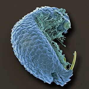
Sperm tails. Transmission electron micrograph (TEM) of sections through the tails of sperm (spermatozoa) cells at various levels. A sperms tails ability to move is partly due to the arrangement of microtubules (red central dots) within the tail. The microtubules are arranged in a central axoneme with two central single microtubules and nine peripheral double microtubules. Surrounding the microtubules are dense fibres, which provide rigidity and protection to the tail. Next is a layer of mitochondria (green) which provide the energy for movement. Finally the tail is wrapped in a protective outer sheath. Magnification: x45, 000 when printed 10 centimetres wide
Science Photo Library features Science and Medical images including photos and illustrations
Media ID 9227051
© STEVE GSCHMEISSNER/SCIENCE PHOTO LIBRARY
Accessory Axoneme Cell Biology Central Cytological Cytology Dense Fibre Fibres Flagella Flagellum Gamete Gametes Microtubule Microtubules Mitochondria Mitochondrion Reproduction Reproductive Sections Sex Cell Sheath Sperm Spermatozoa Spermatozoan Spermatozoon Tail Tails Transmission Electron Micrograph Transmission Electron Microscope Cutouts Section Sectioned
EDITORS COMMENTS
This print showcases the intricate structure of sperm tails, revealing the remarkable mechanisms behind their ability to move. Taken using a transmission electron microscope (TEM), this image provides a close-up view of sections through the tails of sperm cells at various levels. The red central dots in the micrograph represent microtubules, which play a crucial role in facilitating movement. These microtubules are arranged in a central axoneme consisting of two single microtubules and nine double microtubules on the periphery. Surrounding them are dense fibers that provide rigidity and protection to the tail. Adding to its complexity, a layer of green mitochondria can be observed next to these fibers. These mitochondria serve as powerhouses, supplying energy for the sperm's movement. Finally, wrapping around all these components is a protective outer sheath. With magnification at x45,000 when printed 10 centimeters wide, this image offers an extraordinary glimpse into the biological wonders within our bodies. It highlights not only the anatomical features but also emphasizes how vital each component is for successful reproduction. Photographed against a white background with precision and clarity by Steve Gschmeissner from Science Photo Library, this print captures both scientific beauty and educational value. Whether used for research purposes or simply admired for its aesthetic appeal, this photograph invites viewers into an unseen world where life's mysteries unfold before our eyes.
MADE IN AUSTRALIA
Safe Shipping with 30 Day Money Back Guarantee
FREE PERSONALISATION*
We are proud to offer a range of customisation features including Personalised Captions, Color Filters and Picture Zoom Tools
SECURE PAYMENTS
We happily accept a wide range of payment options so you can pay for the things you need in the way that is most convenient for you
* Options may vary by product and licensing agreement. Zoomed Pictures can be adjusted in the Cart.