Fraud Blocker Skip to main content

Cytological Collection

"Cytological Wonders Unveiled: Exploring the Intricacies of Cellular Life" In this captivating journey into the microscopic world, we delve into the realm of cytology

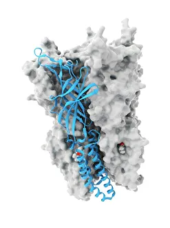
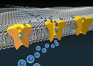
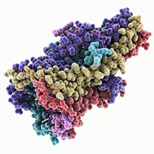
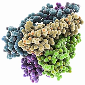
Background imageCytological Collection: Anaesthetic inhibiting an ion channel C015 / 6718

Anaesthetic inhibiting an ion channel C015 / 6718
Anaesthetic inhibiting an ion channel. Computer model showing the structure of propofol anaesthetic drug molecules (spheres)

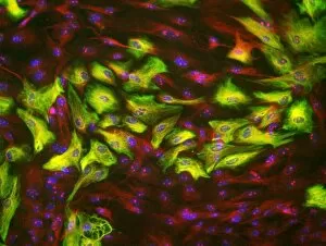
Background imageCytological Collection: HeLa cells, light micrograph C017 / 8299

HeLa cells, light micrograph C017 / 8299
HeLa cells, multiphoton fluorescence micrograph (MFM). The cell nuclei, which contain the cells genetic information, are purple. Microtubules are blue and actin microfilaments are red

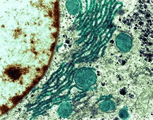
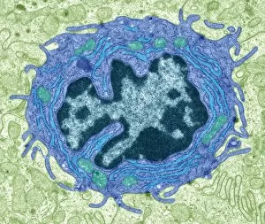
Background imageCytological Collection: Rough endoplasmic reticulum, TEM

Rough endoplasmic reticulum, TEM
Rough endoplasmic reticulum, coloured transmission electron micrograph (TEM). This section shows the rough endoplasmic reticulum (ER, folds, centre), a membranous structure that occurs in cells

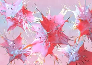
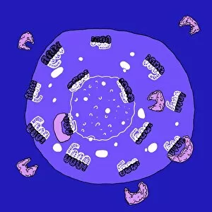
Background imageCytological Collection: Dendritic cells, artwork

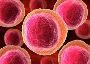
Dendritic cells, artwork. Dendritic cells, a type of white blood cell, are called antigen presenting cells (APCs) and are found in the bodys tissues

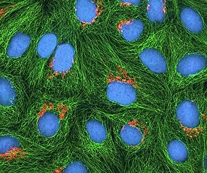
Background imageCytological Collection: HeLa cells, light micrograph C017 / 8298

HeLa cells, light micrograph C017 / 8298
HeLa cells, multiphoton fluorescence micrograph (MFM). The cell nuclei, which contain the cells genetic information, are blue. Golgi bodies, which modify and package proteins, are orange

Background imageCytological Collection: Mitosis, light micrograph

Mitosis, light micrograph
Mitosis. Confocal light micrograph of the stages of mitosis (nuclear division) and cytokinesis (cell division). During mitosis the nuclear envelope disintegrates (3rd image) and the chromosomes (blue)

Background imageCytological Collection: Pine pollen grains, light micrograph

Pine pollen grains, light micrograph
Pine pollen grains. Light micrograph of a section through winged pollen grains (microspores) from a pine (Pinus sp.) tree

Background imageCytological Collection: Pine stem, light micrograph

Pine stem, light micrograph
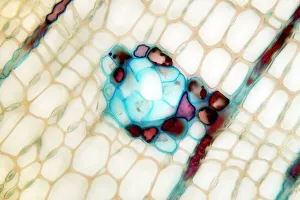
Pine stem. Light micrograph of a section through the stem of a pine (Pinus sp.) tree, showing xylem tissue. The xylem is made up of tracheid cells (light pink)

Background imageCytological Collection: Lime tree stem, light micrograph

Lime tree stem, light micrograph
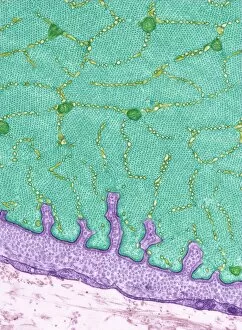
Lime tree stem. Light micrograph of a section through the stem of a lime tree (Tilia europaea). The outer epidermis has been shed and replaced by a layer of cork (dark red)

Background imageCytological Collection: Cell division, fluorescent micrograph

Cell division, fluorescent micrograph
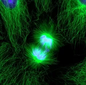
Cell division. Immunofluorescent light micrograph of a human epithelial cell (centre) during the late anaphase stage of mitosis

Background imageCytological Collection: Activated macrophage, SEM C015 / 6375

Activated macrophage, SEM C015 / 6375
Activated macrophage. Coloured scanning electron micrograph (SEM) of an active macrophage white blood cell. Macrophages are cells of the bodys immune system that are found in the tissues rather than

Background imageCytological Collection: Dividing cells

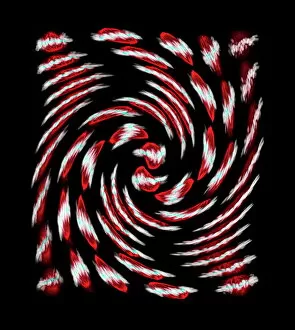
Dividing cells. Computer artwork of a spiral of cells during mitosis (nuclear division). During mitosis two daughter nuclei are formed from one parent nucleus

Background imageCytological Collection: Cell division, fluorescent micrograph

Cell division, fluorescent micrograph
Cell division. Immunofluorescent light micrograph of a human epithelial cell (centre) during the interphase stage of mitosis

Background imageCytological Collection: Ovarian follicle, light micrograph C016 / 0519

Ovarian follicle, light micrograph C016 / 0519
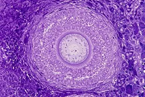
Ovarian follicle. Light micrograph of a section through a secondary ovarian follicle showing signs of maturation into the antral follicle class

Background imageCytological Collection: Eye muscle, TEM C014 / 1468

Eye muscle, TEM C014 / 1468
Eye muscle. Transmission electron micrograph (TEM) of a section through a striated muscle cell from the ciliary muscle of a human eye

Background imageCytological Collection: Purkinje nerve cell, TEM C014 / 0583

Purkinje nerve cell, TEM C014 / 0583
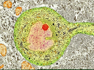
Purkinje nerve cell. Transmission electron micrograph (TEM) of a purkinje nerve cell (green) from the cerebellum of the brain, showing the cell body (centre) and its primary dendrite (cell process)

Background imageCytological Collection: Lymphocyte white blood cells, artwork

Lymphocyte white blood cells, artwork
Lymphocyte white blood cells. Computer artwork of lymphocyte white blood cells, showing their nuclei (red, centre). Lymphocytes, like all white blood cells, are part of the immune system

Background imageCytological Collection: Cell, illustration

Cell, illustration
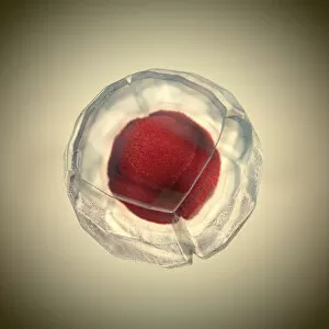
3d illustration of cell division, cell membrane and a splitting red nucleus

Background imageCytological Collection: Cell membrane ion channels, artwork C016 / 7689

Cell membrane ion channels, artwork C016 / 7689
Cell membrane ion channels. Computer artwork of a section through the membrane of an animal cell, showing transmembrane ion channel proteins (yellow)

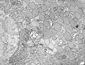
Background imageCytological Collection: Pancreatic acinar cell

Pancreatic acinar cell. Transmission electron micrograph (TEM) of a section through an enzyme-secreting acinar cell in the human pancreas, showing part of the nucleus (round, far left)

Background imageCytological Collection: Hepatocyte liver cell, TEM

Hepatocyte liver cell, TEM
Hepatocyte liver cell. Transmission electron micrograph (TEM) of a section through an hepatocyte liver cell, showing rough and smooth endoplasmic reticulum (ER, dark lines)

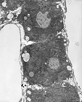
Background imageCytological Collection: Liver tissue, TEM

Liver tissue, TEM
Liver tissue. Transmission electron micrograph (TEM) of a section through the liver, showing part of a radial cord of hepatocyte liver cells (dark) and the vascular sinusoids (white)

Background imageCytological Collection: Cartilage cell, TEM C014 / 1433

Cartilage cell, TEM C014 / 1433
Cartilage cell. Transmission electron micrograph (TEM) of a section through a chondrocyte cell from hyaline cartilage of the trachea (windpipe)

Background imageCytological Collection: Cartilage cell, TEM C014 / 1434

Cartilage cell, TEM C014 / 1434
Cartilage cell. Transmission electron micrograph (TEM) of a section through a chondrocyte cell from hyaline cartilage of the trachea (windpipe)

Background imageCytological Collection: MscL ion channel protein structure

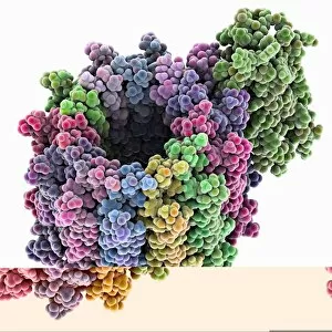
MscL ion channel protein structure. Molecular model showing the protein structure of a Mechanosensitive Channel of Large Conductance (MscL) from a Mycobacterium tuberculosis bacterium

Background imageCytological Collection: Skin cancer cell, SEM C016 / 4764

Skin cancer cell, SEM C016 / 4764
Skin cancer cell. Coloured ion-abrasion scanning electron micrograph (IA-SEM) of a melanoma cell. The large dark sphere is the cell nucleus, which contains the cells genetic information

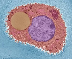
Background imageCytological Collection: Distended fat cell, SEM C013 / 5015

Distended fat cell, SEM C013 / 5015
Distended fat cell. Coloured scanning electron micrograph (SEM) of a large adipocyte (fat cell, orange) distended by fat droplets (green)

Background imageCytological Collection: Distended fat cell, SEM C013 / 5013

Distended fat cell, SEM C013 / 5013
Distended fat cell. Coloured scanning electron micrograph (SEM) of a large adipocyte (fat cell, blue) distended by fat droplets (purple)

Background imageCytological Collection: Osteoblast bone cell, SEM

Osteoblast bone cell, SEM
Osteoblast bone cell. Coloured scanning electron micrograph (SEM) of an osteoblast (centre) in bone tissue. Osteoblasts are primarily found in regions of new bone growth

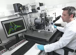
Background imageCytological Collection: Cell biology laboratory

Cell biology laboratory. Scientist using confocal fluorescence microscopy to view stem cells

Background imageCytological Collection: Stochastic gene expression, illustration C018 / 0906

Stochastic gene expression, illustration C018 / 0906
Stochastic gene expression, illustration. Every cell in an organism contains every single gene that makes up the organisms genome. However, they are not all active (expressed) in each cell

Background imageCytological Collection: Cancer cell, artwork C016 / 9843

Cancer cell, artwork C016 / 9843
Cancer cell. Computer artwork of a cancerous (malignant) cell (top left) in tissue. The uneven surface and cytoplasmic projections are characteristic of cancer cells

Background imageCytological Collection: Mitochondria, artwork

Mitochondria, artwork
Mitochondria, computer artwork

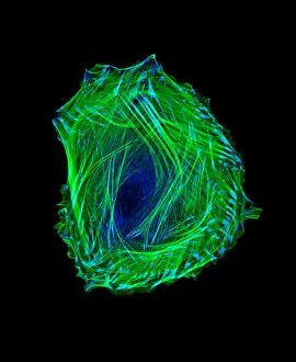
Background imageCytological Collection: Embryonic smooth muscle cell C018 / 8595

Embryonic smooth muscle cell C018 / 8595
Embryonic smooth muscle cell, immunofluorescence micrograph. Actin filaments are green, the protein vinculin is light blue. Actin is part of the cytoskeleton

Background imageCytological Collection: Eosinophil white blood cell, TEM C014 / 1438

Eosinophil white blood cell, TEM C014 / 1438
Eosinophil white blood cell. Transmission electron micrograph (TEM) of a section through an eosinophil. Eosinophils, like all white blood cells, are part of the bodys immune system

Background imageCytological Collection: Bladder lining, TEM C014 / 1473

Bladder lining, TEM C014 / 1473
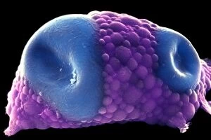
Bladder lining. Transmission electron micrograph (TEM) of a section through the epithelial cells that line of a human bladder, showing large flat vesicles (large, blue)

Background imageCytological Collection: Intestinal gland cell, TEM C014 / 1441

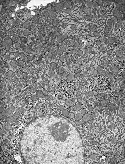
Intestinal gland cell, TEM C014 / 1441
Intestinal gland cell. Transmission electron micrograph (TEM) of a section through an undifferentiated cell from the intestinal tract, showing extensive rough endoplasmic reticulum (ER)

Background imageCytological Collection: Potassium ion channel protein structure

Potassium ion channel protein structure. Molecular model of a KcsA potassium ion (K+) channel from Streptomyces lividans bacteria

Background imageCytological Collection: Potassium ion channel beta subunit

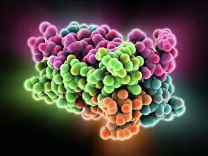
Potassium ion channel beta subunit. Molecular model showing the structure a beta subunit of a voltage-dependent potassium (K+) channel

Background imageCytological Collection: KCNQ ion channel protein structure

KCNQ ion channel protein structure. Molecular model showing the protein structure of an ion channel domain. Ion channels are membrane-spanning proteins that form a pathway for the movement of

Background imageCytological Collection: Potassium ion channel cavity structure

Potassium ion channel cavity structure. Molecular model showing the structure of a cavity formed by potassium ion channel proteins

Background imageCytological Collection: ATP synthase molecule C014 / 0880

ATP synthase molecule C014 / 0880
ATP synthase molecule. Molecular model showing the structure of ATP synthase (ATPase) subunit C. ATPase is an important enzyme that provides energy for cells through the synthesis of adenosine

Background imageCytological Collection: Astrocyte brain cells, light micrograph

Astrocyte brain cells, light micrograph
Astrocyte brain cells. Fluorescence light micrograph of primary astrocyte cells from the brain of a mouse. Astrocytes have numerous branches of connective tissue that provide support

Background imageCytological Collection: HeLa cell, SEM C014 / 0371

HeLa cell, SEM C014 / 0371
HeLa cell. Coloured scanning electron micrograph (SEM) of a HeLa cell (centre) grown in a 3D matrix (background). HeLa cells are a continuously cultured cell line of immortal human cancer cells

Background imageCytological Collection: Intestinal gland cell, TEM C014 / 1440

Intestinal gland cell, TEM C014 / 1440
Intestinal gland cell. Transmission electron micrograph (TEM) of a section through an undifferentiated cell from the intestinal tract, showing extensive rough endoplasmic reticulum (ER)

Background imageCytological Collection: Cartilage cell, TEM C014 / 1432

Cartilage cell, TEM C014 / 1432
Cartilage cell. Transmission electron micrograph (TEM) of a section through a chondrocyte cell from hyaline cartilage of the trachea (windpipe)

Background imageCytological Collection: Wnt signalling pathways, illustration C018 / 0917

Wnt signalling pathways, illustration C018 / 0917
Wnt signalling pathways, illustration. Wnt signalling pathways are three separate pathways that pass signals from outside a cell to inside the cell

Background imageCytological Collection: Fat cells, SEM C014 / 0364

Fat cells, SEM C014 / 0364
Fat cells. Coloured scanning electron micrograph (SEM) of adipocytes (fat cells). Adipocytes store energy as an insulating layer of fat


All Professionally Made to Order for Quick Shipping

"Cytological Wonders Unveiled: Exploring the Intricacies of Cellular Life" In this captivating journey into the microscopic world, we delve into the realm of cytology, where astonishing discoveries await. From anaesthetic inhibiting an ion channel to mesmerizing light and electron micrographs, each image offers a glimpse into the intricate workings of cells. Firstly, we witness an extraordinary phenomenon as anaesthetic molecules inhibit an ion channel within cells. This interaction sheds light on how these substances affect cellular processes and highlights their potential therapeutic applications. Moving forward, our attention is drawn to HeLa cells captured under a light microscope. The vivid details reveal their complex structures and provide valuable insights for research in various fields such as cancer biology and drug development. Next up is a stunning transmission electron micrograph showcasing the rough endoplasmic reticulum. Its labyrinthine network serves as a hub for protein synthesis and transport within cells, emphasizing its crucial role in maintaining cellular homeostasis. As we shift gears towards artistic representation, dendritic cells take center stage through exquisite artwork. These specialized immune cells play a pivotal role in recognizing foreign invaders and orchestrating immune responses - truly nature's defenders at work. Returning to HeLa cells under another light microscope lens unveils yet another breathtaking display of cellular beauty. Each cell appears like a universe unto itself with intricate organelles working harmoniously to sustain life's delicate balance. The enchantment continues with captivating images capturing mitosis - the process by which one cell divides into two identical daughter cells. Witnessing this dance of chromosomes during cell division provides profound insights into growth, development, and regeneration mechanisms within organisms. Shifting focus from animal to plant life brings us pine pollen grains delicately observed under a light microscope. These tiny particles hold immense significance in plant reproduction dynamics while exhibiting remarkable structural intricacies that aid successful pollination. Further exploring plants' hidden wonders takes us on an enlightening journey through light micrographs of pine and lime tree stems.