Fraud Blocker Skip to main content

Cytological Collection (page 5)

"Cytological Wonders Unveiled: Exploring the Intricacies of Cellular Life" In this captivating journey into the microscopic world, we delve into the realm of cytology

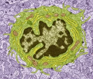
Background imageCytological Collection: Dividing cell, TEM

Dividing cell, TEM
Dividing cell. Transmission electron micrograph (TEM) of a section through a cell dividing through the process of mitosis

Background imageCytological Collection: Myelinated nerve tissue, TEM

Myelinated nerve tissue, TEM
Myelinated nerve tissue. Transmission electron micrograph (TEM) of a section through myelinated nerve fibres (axons). Each axon is coated with many layers of myelin

Background imageCytological Collection: Ovarian follicle, TEM

Ovarian follicle, TEM
Ovarian follicle. Transmission electron micrograph (TEM) of a section through an ovary, showing a primary follicle. Primary follicles contain a central oocyte (female germ cell)

Background imageCytological Collection: Smooth muscle tissue, TEM

Smooth muscle tissue, TEM
Smooth muscle tissue. Transmission electron micrograph (TEM) of a longitudinal section through smooth muscle cells. Smooth muscle cells are spindle-shaped and often closely packed together

Background imageCytological Collection: Anterior pituitary gland, TEM

Anterior pituitary gland, TEM
Anterior pituitary gland. Transmission electron micrograph (TEM) of a section through cells of the anterior pituitary. The anterior pituitary, or adenohypophysis

Background imageCytological Collection: Enteroendocrine cell, TEM

Enteroendocrine cell, TEM
Enteroendocrine cell. Transmission electron micrograph (TEM) of a section through an enteroendocrine cell within a gland of the stomach

Background imageCytological Collection: Cyclin-dependent kinase inhibitor complex

Cyclin-dependent kinase inhibitor complex. Molecular model of the inhibitor protein P27Kip1 bound to a cyclin-A-cyclin dependent kinase 2 complex

Background imageCytological Collection: Antibodies and B cells, artwork C016 / 6255

Antibodies and B cells, artwork C016 / 6255
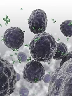
Antibodies and B cells. Computer artwork showing antibodies (green) neutralising cytokine activity in B cell (large, round) white blood cells

Background imageCytological Collection: Antibodies and B cells, artwork C016 / 6254

Antibodies and B cells, artwork C016 / 6254
Antibodies and B cells. Computer artwork showing antibodies (green) neutralising cytokine activity in B cell (large, round) white blood cells

Background imageCytological Collection: Drug effect on viruses, conceptual image C016 / 6253

Drug effect on viruses, conceptual image C016 / 6253
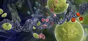
Drug effect on viruses, conceptual image. Computer artwork showing a single strand of DNA (deoxyribonucleic acid, spiral, centre), red blood cells (pink), virus particles (virions, green, small)

Background imageCytological Collection: Prokaryote cell, artwork C016 / 6263

Prokaryote cell, artwork C016 / 6263
Prokaryote cell. Computer artwork showing the internal structure of a typical prokaryotic cell. Prokaryotes are organisms whose cells lack a membrane-bound nucleus (karyon)

Background imageCytological Collection: Eukaryote cell, artwork C016 / 6260

Eukaryote cell, artwork C016 / 6260
Eukaryote cell. Computer artwork showing the internal structure of a typical eukaryotic cell. Eukaryotes are organisms whose cells contain a membrane-bound nucleus (karyon, orange)

Background imageCytological Collection: Prokaryote and eukaryote cells, artwork C016 / 6259

Prokaryote and eukaryote cells, artwork C016 / 6259
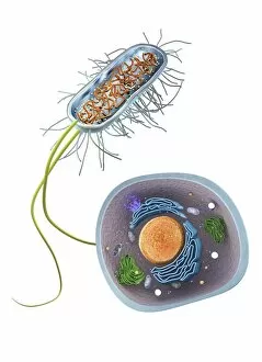
Prokaryote and eukaryote cells. Computer artwork comparing typical prokaryotic (top) and eukaryotic (bottom) cells. Prokaryotes are organisms whose cells lack a membrane-bound nucleus (karyon)

Background imageCytological Collection: Effect of oxygen supply on tumour growth C016 / 6256

Effect of oxygen supply on tumour growth C016 / 6256
Effect of oxygen supply on tumour growth. Top: a small blood supply (tubes) means a low level of oxygen is available and the tumour (round) grows slowly

Background imageCytological Collection: Gastritis, light micrograph C016 / 0508

Gastritis, light micrograph C016 / 0508
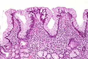
Gastritis. Light micrograph of a section through an inflamed human stomach (gastritis), showing an abnormal accumulation of white blood cells (leucocytes)

Background imageCytological Collection: Corpus luteum of ovary, light micrograph C016 / 0527

Corpus luteum of ovary, light micrograph C016 / 0527
Corpus luteum of ovary. Light micrograph of a section through steroid-secreting luteal cells in the corpus luteum of an ovary. The corpus luteum develops from an ovulated follicle

Background imageCytological Collection: Appendix, light micrograph C016 / 0510

Appendix, light micrograph C016 / 0510
Appendix. Light micrograph of a section through a human appendix. The appendix is a blind-ending small diameter tubular extension of the gut, arising from the caecum of the large bowel

Background imageCytological Collection: Retina of the eye, light micrograph C016 / 0528

Retina of the eye, light micrograph C016 / 0528
Retina of the eye. Light micrograph of a section through the retina from a human eye. From top down: nerve fibres of the optic nerve and a blood vessel; several layers of neurons (nerve cells)

Background imageCytological Collection: Gastritis, light micrograph C016 / 0507

Gastritis, light micrograph C016 / 0507
Gastritis. Light micrograph of a section through an inflamed human stomach (gastritis), showing an abnormal accumulation of white blood cells (leucocytes)

Background imageCytological Collection: Cancellous bone, light micrograph C016 / 0509

Cancellous bone, light micrograph C016 / 0509
Cancellous bone. Light micrograph of a section through cancellous, or spongy, bone. Constructed in a honeycomb-type architecture

Background imageCytological Collection: Colon polyp, light micrograph C016 / 0512

Colon polyp, light micrograph C016 / 0512
Colon polyp. Light micrograph of a section through part of a tubulovillous adenoma of the mucosa of the large bowel (colon)

Background imageCytological Collection: Lens of the eye, light micrograph C016 / 0526

Lens of the eye, light micrograph C016 / 0526
Lens of the eye. Light micrograph of a section through the lens from a human eye. The lens consists of thousands of precisely aligned stacks of fibres seen here end-on

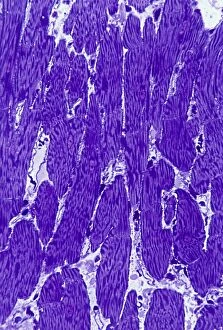
Background imageCytological Collection: Heart muscle, light micrograph C016 / 0517

Heart muscle, light micrograph C016 / 0517
Heart muscle. Light micrograph of a section through heart (cardiac) muscle tissue, showing cardiomyocyte muscle cells. These cells have a centrally located nucleus and are branched

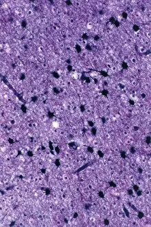
Background imageCytological Collection: Brain glial cells, light micrograph C016 / 0524

Brain glial cells, light micrograph C016 / 0524
Brain glial cells. Light micrograph of a section through brain tissue, showing numerous glial cells (dark). Glial cells, or neuroglia, are non-neural cells of the central nervous system (CNS)

Background imageCytological Collection: Cirrhosis of liver, light micrograph C016 / 0530

Cirrhosis of liver, light micrograph C016 / 0530
Cirrhosis of liver. Light micrograph of a section through liver tissue, damaged by cirrhosis. Cirrhosis occurs as a result of a range of factors causing damage to liver function

Background imageCytological Collection: Stratified squamous epithelium C016 / 0534

Stratified squamous epithelium C016 / 0534
Stratified squamous epithelium. Light micrograph of a section through stratified squamous epithelium, showing the multiple layers, or strata, that function to resist abrasion of the surface

Background imageCytological Collection: Nerve ganglion, light micrograph C016 / 0532

Nerve ganglion, light micrograph C016 / 0532
Nerve ganglion. Light micrograph of a section through a nerve ganglion of the peripheral nervous system, showing clusters of nerve cell bodies

Background imageCytological Collection: Myelinated nerve fibres, light micrograph C016 / 0531

Myelinated nerve fibres, light micrograph C016 / 0531
Myelinated nerve fibres. Light micrograph of a section through a spinal sensory nerve containing many closely packed axons exhibiting a wave-type appearance

Background imageCytological Collection: Colon, light micrograph C016 / 0516

Colon, light micrograph C016 / 0516
Colon. Light micrograph of a section through the mucosa (inner epithelial lining) of the colon (large bowel), showing the typical arrangement of invaginations of columnar epithelial cells forming

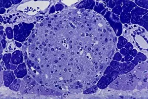
Background imageCytological Collection: Islet of Langerhans, light micrograph C016 / 0514

Islet of Langerhans, light micrograph C016 / 0514
Islet of Langerhans. Light micrograph of a section through an islet of Langerhans in a pancreas. Scattered throughout the pancreas, the islets are endocrine cells that secrete a range of hormones

Background imageCytological Collection: Bowel cancer, light micrograph C016 / 0511

Bowel cancer, light micrograph C016 / 0511
Bowel cancer. Light micrograph of a section through a carcinoid tumour in the small bowel. The tumour has invaded the submucosa of the bowel

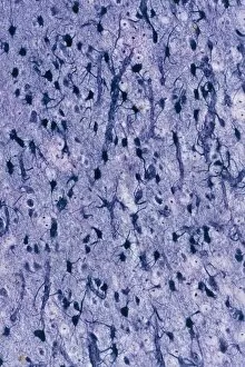
Background imageCytological Collection: Brain glial cells, light micrograph C016 / 0523

Brain glial cells, light micrograph C016 / 0523
Brain glial cells. Light micrograph of a section through brain tissue, showing numerous glial cells (dark). Glial cells, or neuroglia, are non-neural cells of the central nervous system (CNS)

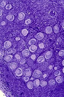
Background imageCytological Collection: Ovarian primordial follicles, micrograph C016 / 0518

Ovarian primordial follicles, micrograph C016 / 0518
Ovarian primordial follicles. Light micrograph of a section through the cortex of a neonatal ovary, showing clusters of primordial follicles each containing a single oocyte (egg cell)

Background imageCytological Collection: Islet of Langerhans, light micrograph C016 / 0513

Islet of Langerhans, light micrograph C016 / 0513
Islet of Langerhans. Light micrograph of a section through an islet of Langerhans in a pancreas. Scattered throughout the pancreas, the islets are endocrine cells that secrete a range of hormones

Background imageCytological Collection: Cirrhosis of liver, light micrograph C016 / 0529

Cirrhosis of liver, light micrograph C016 / 0529
Cirrhosis of liver. Light micrograph of a section through liver tissue, damaged by cirrhosis. Cirrhosis occurs as a result of a range of factors causing damage to liver function

Background imageCytological Collection: Neutrophil white blood cell, illustration C018 / 0741

Neutrophil white blood cell, illustration C018 / 0741
Neutrophil white blood cell, illustration. Neutrophils are the most abundant white blood cell in humans. They have a multi-lobed nucleus (dark purple)

Background imageCytological Collection: Meiosis, illustration C018 / 0803

Meiosis, illustration C018 / 0803
Meiosis. Illustration of a cell during metaphase (I) of meiosis (gamete formation). During meiosis four daughter nuclei are formed from one parent nucleus after two stages of nuclear division

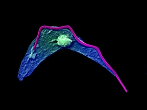
Background imageCytological Collection: Cytoskeleton in unicellular parasite, SEM C018 / 0518

Cytoskeleton in unicellular parasite, SEM C018 / 0518
Cytoskeleton in unicellular parasite, coloured scanning electron micrograph (SEM). All cells have a support and transport network called the cytoskeleton

Background imageCytological Collection: Gene expression, conceptual illustration C018 / 0746

Gene expression, conceptual illustration C018 / 0746
Gene expression, conceptual illustration. Every cell in an organism contains every single gene that makes up the organisms genome. However, they are not all active (expressed) in each cell

Background imageCytological Collection: Animal cell, illustration C018 / 0734

Animal cell, illustration C018 / 0734
Animal cell. Illustration showing the organelles present in an animal cell. At lower centre is the nucleus, which contains the cells genetic information in the form of DNA (deoxyribonucleic acid)

Background imageCytological Collection: Mechanical stress on a cell, illustration C018 / 0776

Mechanical stress on a cell, illustration C018 / 0776
Mechanical stress on a cell, conceptual illustration. Mechanical stress can have an effect on a number of processes within a cell, including polarity, differentiation, growth and migration

Background imageCytological Collection: Cellular polarity, illustration C018 / 0775

Cellular polarity, illustration C018 / 0775
Cellular Polarity. Conceptual illustration of cell polarity research and its medical implications

Background imageCytological Collection: Microtubule formation, illustration C018 / 0804

Microtubule formation, illustration C018 / 0804
Microtubule formation, illustration. Microtubules are polymers of the protein tubulin and are a component of the cytoskeleton

Background imageCytological Collection: Migrating cell, illustration C018 / 0754

Migrating cell, illustration C018 / 0754
Migrating cell. Time-lapse illustration of a migrating cell, using outlines to show the previous cell boundaries. Cell migration is key to metastasis (spread) and the progression of cancer

Background imageCytological Collection: Eye muscle, TEM C014 / 1467

Eye muscle, TEM C014 / 1467
Eye muscle. Transmission electron micrograph (TEM) of a section through a striated muscle cell from the ciliary muscle of a human eye

Background imageCytological Collection: Intestinal gland cell, TEM C014 / 1442

Intestinal gland cell, TEM C014 / 1442
Intestinal gland cell. Transmission electron micrograph (TEM) of a section through an undifferentiated cell from the intestinal tract, showing extensive rough endoplasmic reticulum (ER)

Background imageCytological Collection: Trachea lining, TEM C014 / 1469

Trachea lining, TEM C014 / 1469
Trachea lining. Transmission electron micrograph (TEM) of a longitudinal section through the lining of the trachea (windpipe), which links the larynx (voicebox) to the lungs

Background imageCytological Collection: Mouth cancer cell, SEM C014 / 1460

Mouth cancer cell, SEM C014 / 1460
Mouth cancer cell. Coloured scanning electron micrograph (SEM) of a squamous cell carcinoma (cancer) cell from a human mouth


All Professionally Made to Order for Quick Shipping

"Cytological Wonders Unveiled: Exploring the Intricacies of Cellular Life" In this captivating journey into the microscopic world, we delve into the realm of cytology, where astonishing discoveries await. From anaesthetic inhibiting an ion channel to mesmerizing light and electron micrographs, each image offers a glimpse into the intricate workings of cells. Firstly, we witness an extraordinary phenomenon as anaesthetic molecules inhibit an ion channel within cells. This interaction sheds light on how these substances affect cellular processes and highlights their potential therapeutic applications. Moving forward, our attention is drawn to HeLa cells captured under a light microscope. The vivid details reveal their complex structures and provide valuable insights for research in various fields such as cancer biology and drug development. Next up is a stunning transmission electron micrograph showcasing the rough endoplasmic reticulum. Its labyrinthine network serves as a hub for protein synthesis and transport within cells, emphasizing its crucial role in maintaining cellular homeostasis. As we shift gears towards artistic representation, dendritic cells take center stage through exquisite artwork. These specialized immune cells play a pivotal role in recognizing foreign invaders and orchestrating immune responses - truly nature's defenders at work. Returning to HeLa cells under another light microscope lens unveils yet another breathtaking display of cellular beauty. Each cell appears like a universe unto itself with intricate organelles working harmoniously to sustain life's delicate balance. The enchantment continues with captivating images capturing mitosis - the process by which one cell divides into two identical daughter cells. Witnessing this dance of chromosomes during cell division provides profound insights into growth, development, and regeneration mechanisms within organisms. Shifting focus from animal to plant life brings us pine pollen grains delicately observed under a light microscope. These tiny particles hold immense significance in plant reproduction dynamics while exhibiting remarkable structural intricacies that aid successful pollination. Further exploring plants' hidden wonders takes us on an enlightening journey through light micrographs of pine and lime tree stems.