Fraud Blocker Skip to main content

Cytological Collection (#3)

"Cytological Wonders Unveiled: Exploring the Intricacies of Cellular Life" In this captivating journey into the microscopic world, we delve into the realm of cytology

Background imageCytological Collection: Myelinated nerve, light micrograph

Myelinated nerve, light micrograph
Myelinated nerve. Light micrograph of a section through a peripheral myelinated nerve, showing many individual axons each covered with deep blue-staining myelin sheaths

Background imageCytological Collection: Kinesin motor protein

Kinesin motor protein. Molecular model of the ncd kinesin motor protein. Kinesin motor proteins transport vesicles containing intracellular cargo around the cell along microtubules

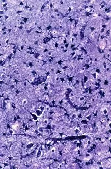
Background imageCytological Collection: Glial cells, light micrograph

Glial cells, light micrograph
Glial cells in the brain. Light micrograph of a section through glial cells (dark) in the grey matter of the brain. Due to their star shape these glial cells are called astrocytes

Background imageCytological Collection: Phospholipid molecule, artwork

Phospholipid molecule, artwork
Phospholipid molecule. Computer artwork showing the structure of a phospholipid molecule. Phospholipids consist of a phosphate group head (top) and a fatty acid hydrophobic ( water-hating ) tail

Background imageCytological Collection: Haematopoietic stem cells, artwork

Haematopoietic stem cells, artwork
Haematopoietic stem cells. Cutaway computer artwork showing white blood cells (leucocytes, white, round), red blood cells (erythrocytes, red) and haematopoietic stem cells (HSCs)

Background imageCytological Collection: Algae cell wall, SEM

Algae cell wall, SEM
Algae cell wall. Coloured scanning electron micrograph (SEM) of cellulose microfibrils (strands) in a Chaetomorpha sp. algal cell wall

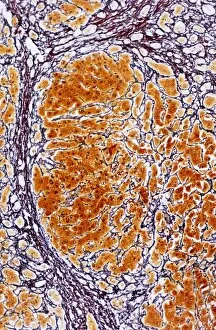
Background imageCytological Collection: Cirrhosis of the liver, light micrograph

Cirrhosis of the liver, light micrograph
Cirrhosis of the liver. Light micrograph of a section through liver tissue affected by cirrhosis, showing a cluster of hepatocyte cells (orange)

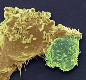
Background imageCytological Collection: White blood cell antigen presentation C016 / 9058

White blood cell antigen presentation C016 / 9058
White blood cell antigen presentation. Coloured scanning electron micrograph (SEM) showing the interaction between a macrophage (yellow) and a T helper lymphocyte (Th cell, green)

Background imageCytological Collection: E. coli bacteria, SEM C016 / 9128

E. coli bacteria, SEM C016 / 9128
E. coli bacteria. Coloured scanning electron micrograph (SEM) of Escherichia coli bacteria (purple) attached to the remains of a dead cell. E

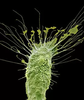
Background imageCytological Collection: Microglial white blood cell, SEM C016 / 9115

Microglial white blood cell, SEM C016 / 9115
Microglial white blood cell. Coloured scanning electron micrograph (SEM) of a microglial cell. Microglia are phagocytic macrophage white blood cells that are the main form of immune system in

Background imageCytological Collection: E. coli bacteria, SEM C016 / 9130

E. coli bacteria, SEM C016 / 9130
E. coli bacteria. Coloured scanning electron micrograph (SEM) of Escherichia coli bacteria (red) attached to the remains of a dead cell. E

Background imageCytological Collection: Microglial white blood cell, SEM C016 / 9119

Microglial white blood cell, SEM C016 / 9119
Microglial white blood cell. Coloured scanning electron micrograph (SEM) of a microglial cell. Microglia are phagocytic macrophage white blood cells that are the main form of immune system in

Background imageCytological Collection: Microglial white blood cell, SEM C016 / 9117

Microglial white blood cell, SEM C016 / 9117
Microglial white blood cell. Coloured scanning electron micrograph (SEM) of a microglial cell. Microglia are phagocytic macrophage white blood cells that are the main form of immune system in

Background imageCytological Collection: Platelet, SEM C016 / 9060

Platelet, SEM C016 / 9060
Platelet, coloured scanning electron micrograph (SEM). Platelets, or thrombocytes, are fragments of white blood cells. When blood clots

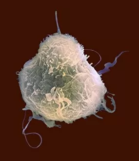
Background imageCytological Collection: Lymphocyte white blood cell, SEM C016 / 9414

Lymphocyte white blood cell, SEM C016 / 9414
Lymphocyte white blood cell. Coloured scanning electron micrograph (SEM) of a lymphocyte white blood cell. Lymphocytes are involved in the immune systems defence mechanisms, lymph system

Background imageCytological Collection: E. coli bacteria, SEM C016 / 9127

E. coli bacteria, SEM C016 / 9127
E. coli bacteria. Coloured scanning electron micrograph (SEM) of Escherichia coli bacteria (green) attached to the remains of a dead cell. E

Background imageCytological Collection: Microglial white blood cell, SEM C016 / 9109

Microglial white blood cell, SEM C016 / 9109
Microglial white blood cell. Coloured scanning electron micrograph (SEM) of a microglial cell. Microglia are phagocytic macrophage white blood cells that are the main form of immune system in

Background imageCytological Collection: Lymphocyte white blood cell, SEM C016 / 9415

Lymphocyte white blood cell, SEM C016 / 9415
Lymphocyte white blood cell. Coloured scanning electron micrograph (SEM) of a lymphocyte white blood cell. Lymphocytes are involved in the immune systems defence mechanisms, lymph system

Background imageCytological Collection: Dicotyledon vascular bundle, SEM C016 / 9603

Dicotyledon vascular bundle, SEM C016 / 9603
Dicotyledon vascular bundle. Coloured scanning electron micrograph (SEM) of a section through a vascular bundle from a dicotyledonous plant

Background imageCytological Collection: Dicotyledon vascular bundle, SEM C016 / 9602

Dicotyledon vascular bundle, SEM C016 / 9602
Dicotyledon vascular bundle. Coloured scanning electron micrograph (SEM) of a section through a vascular bundle from a dicotyledonous plant

Background imageCytological Collection: Roundworm germ cells, light micrograph C016 / 9538

Roundworm germ cells, light micrograph C016 / 9538
Roundworm germ cells. Light micrograph of germ cells from a roundworm (Ascaris sp.), undergoing mitosis (nuclear division)

Background imageCytological Collection: White blood cell antigen presentation C016 / 9057

White blood cell antigen presentation C016 / 9057
White blood cell antigen presentation. Coloured scanning electron micrograph (SEM) showing the interaction between a macrophage (red) and a T helper lymphocyte (Th cell, yellow)

Background imageCytological Collection: Microglial white blood cell, SEM C016 / 9114

Microglial white blood cell, SEM C016 / 9114
Microglial white blood cell. Coloured scanning electron micrograph (SEM) of a microglial cell. Microglia are phagocytic macrophage white blood cells that are the main form of immune system in

Background imageCytological Collection: Microglial white blood cells, SEM C016 / 9113

Microglial white blood cells, SEM C016 / 9113
Microglial white blood cells. Coloured scanning electron micrograph (SEM) of microglial cells. Microglia are phagocytic macrophage white blood cells that are the main form of immune system in

Background imageCytological Collection: Microglial white blood cell, SEM C016 / 9118

Microglial white blood cell, SEM C016 / 9118
Microglial white blood cell. Coloured scanning electron micrograph (SEM) of a microglial cell. Microglia are phagocytic macrophage white blood cells that are the main form of immune system in

Background imageCytological Collection: Microglial white blood cells, SEM C016 / 9111

Microglial white blood cells, SEM C016 / 9111
Microglial white blood cells. Coloured scanning electron micrograph (SEM) of microglial cells. Microglia are phagocytic macrophage white blood cells that are the main form of immune system in

Background imageCytological Collection: Platelet, SEM C016 / 9059

Platelet, SEM C016 / 9059
Platelet, coloured scanning electron micrograph (SEM). Platelets, or thrombocytes, are fragments of white blood cells. When blood clots

Background imageCytological Collection: Microglial white blood cells, SEM C016 / 9112

Microglial white blood cells, SEM C016 / 9112
Microglial white blood cells. Coloured scanning electron micrograph (SEM) of microglial cells. Microglia are phagocytic macrophage white blood cells that are the main form of immune system in

Background imageCytological Collection: Microglial white blood cell, SEM C016 / 9116

Microglial white blood cell, SEM C016 / 9116
Microglial white blood cell. Coloured scanning electron micrograph (SEM) of a microglial cell. Microglia are phagocytic macrophage white blood cells that are the main form of immune system in

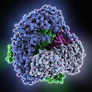
Background imageCytological Collection: Adaptor protein-1 molecule

Adaptor protein-1 molecule
Adaptor protein-1, molecular model. This protein is found in the trans-Golgi network (TGN) of eukaryotic cells, where modified proteins are packaged and sorted for transport around the cell

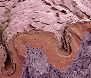
Background imageCytological Collection: Finger skin, SEM

Finger skin, SEM
Finger skin. Coloured scanning electron micrograph (SEM) of a section through skin from a human finger, showing the characteristic dermal ridges (lower left, and right) that make up the fingerprint

Background imageCytological Collection: Corpus luteum, TEM

Corpus luteum, TEM
Corpus luteum. Transmission electron micrograph (TEM) of a section through the corpus luteum of an ovary, showing several luteal cells

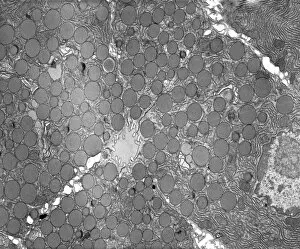
Background imageCytological Collection: Pancreatic exocrine cells, TEM

Pancreatic exocrine cells, TEM
Pancreatic exocrine cells. Transmission electron micrograph (TEM) of a section through exocrine cells in the pancreas, showing numerous zymogen granules (circles), rough endoplasmic reticulum (ER)

Background imageCytological Collection: Glomerulus, TEM

Glomerulus, TEM
Glomerulus. Transmission electron micrograph (TEM) of a section through a nephron in a human kidney, showing a cluster of capillaries within a renal corpuscle, also known as a glomerulus

Background imageCytological Collection: Gastric gland, TEM

Gastric gland, TEM
Gastric gland. Transmission electron micrograph (TEM) of a section through the deep region of a gastric (fundus) gland, showing several enzyme-secreting chief cells with many secretory granules

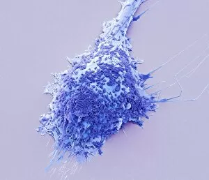
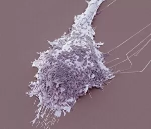
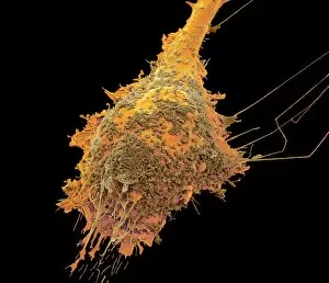
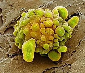
Background imageCytological Collection: Stem cell dying, SEM

Stem cell dying, SEM
Stem cell dying. Coloured scanning electron micrograph (SEM) of a stem cell undergoing apoptosis, or programmed cell death. Apoptosis occurs when a cell becomes old or damaged

Background imageCytological Collection: Islet of Langerhans beta cell, TEM

Islet of Langerhans beta cell, TEM
Islet of Langerhans beta cell. Transmission electron micrograph (TEM) of a section through a beta cell in an islet of Langerhans in the pancreas

Background imageCytological Collection: Pancreatic acinar cell, TEM

Pancreatic acinar cell, TEM
Pancreatic acinar cell. Transmission electron micrograph (TEM) of a section through an enzyme-secreting acinar cell in the human pancreas, showing the nucleus (dark purple, centre)

Background imageCytological Collection: Basophil white blood cell, TEM

Basophil white blood cell, TEM
Basophil white blood cell. Transmission electron micrograph (TEM) of section through a circulating basophil cell. Basophils are the smallest and least common of the white blood cells

Background imageCytological Collection: Myenteric nerve plexus, TEM

Myenteric nerve plexus, TEM
Myenteric nerve plexus. Transmission electron micrograph (TEM) of a section through a bundle of unmyelinated nerve fibres in the myenteric nerve plexus of the intestinal tract

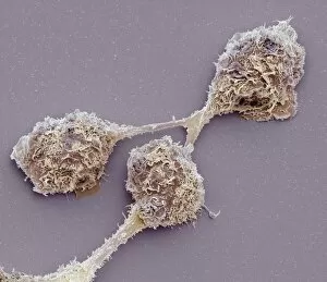
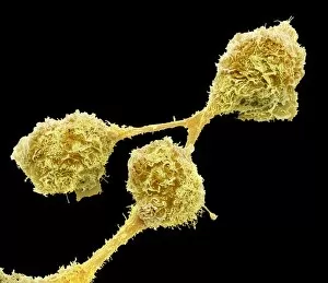
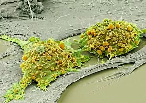
Background imageCytological Collection: Mesenchymal stem cells, SEM

Mesenchymal stem cells, SEM
Mesenchymal stem cells. Coloured scanning electron micrograph (SEM) of two human mesenchymal stem cells (MSCs). MSCs are multipotent stromal (connective tissue)

Background imageCytological Collection: Golgi membranes, TEM

Golgi membranes, TEM
Golgi membranes. Transmission electron micrograph (TEM) of a section through a cell, showing the membranes (dark lines) of the Golgi apparatus

Background imageCytological Collection: Goblet cell, TEM

Goblet cell, TEM
Goblet cell. Transmission electron micrograph (TEM) of a section through a goblet cell in the lining (epithelium, top) of the small intestine

Background imageCytological Collection: Smooth muscle of gut wall, TEM

Smooth muscle of gut wall, TEM
Smooth muscle of gut wall. Transmission electron micrograph (TEM) of a section through the outermost wall of the intestine

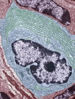
Background imageCytological Collection: Lymphocyte white blood cell, TEM

Lymphocyte white blood cell, TEM
Lymphocyte white blood cell. Transmission electron micrograph (TEM) of a section through lymphocyte, showing the nucleus (large, round, centre), endoplasmic reticulum (ER)

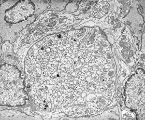
Background imageCytological Collection: Unmyelinated nerve, TEM

Unmyelinated nerve, TEM
Unmyelinated nerve. Transmission electron micrograph (TEM) of a section through axon (nerve fibre) bundles of unmyelinated nerves

Background imageCytological Collection: Steroid secreting cell, TEM

Steroid secreting cell, TEM
Steroid secreting cell. Transmission electron micrograph (TEM) of a section through a cell involved with steroid synthesis and secretion (steroidogenesis)

Background imageCytological Collection: Enterocyte, TEM

Enterocyte, TEM
Enterocyte. Transmission electron micrograph (TEM) of a section through the cytoplasm and part of the nucleus of an enterocyte cell located in a crypt of Lieberkuhn of the small intestine


All Professionally Made to Order for Quick Shipping

"Cytological Wonders Unveiled: Exploring the Intricacies of Cellular Life" In this captivating journey into the microscopic world, we delve into the realm of cytology, where astonishing discoveries await. From anaesthetic inhibiting an ion channel to mesmerizing light and electron micrographs, each image offers a glimpse into the intricate workings of cells. Firstly, we witness an extraordinary phenomenon as anaesthetic molecules inhibit an ion channel within cells. This interaction sheds light on how these substances affect cellular processes and highlights their potential therapeutic applications. Moving forward, our attention is drawn to HeLa cells captured under a light microscope. The vivid details reveal their complex structures and provide valuable insights for research in various fields such as cancer biology and drug development. Next up is a stunning transmission electron micrograph showcasing the rough endoplasmic reticulum. Its labyrinthine network serves as a hub for protein synthesis and transport within cells, emphasizing its crucial role in maintaining cellular homeostasis. As we shift gears towards artistic representation, dendritic cells take center stage through exquisite artwork. These specialized immune cells play a pivotal role in recognizing foreign invaders and orchestrating immune responses - truly nature's defenders at work. Returning to HeLa cells under another light microscope lens unveils yet another breathtaking display of cellular beauty. Each cell appears like a universe unto itself with intricate organelles working harmoniously to sustain life's delicate balance. The enchantment continues with captivating images capturing mitosis - the process by which one cell divides into two identical daughter cells. Witnessing this dance of chromosomes during cell division provides profound insights into growth, development, and regeneration mechanisms within organisms. Shifting focus from animal to plant life brings us pine pollen grains delicately observed under a light microscope. These tiny particles hold immense significance in plant reproduction dynamics while exhibiting remarkable structural intricacies that aid successful pollination. Further exploring plants' hidden wonders takes us on an enlightening journey through light micrographs of pine and lime tree stems.