Fraud Blocker Skip to main content

Mitochondria Collection

Mitochondria, the powerhouse of the cell, play a vital role in various organisms

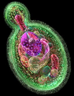
Background imageMitochondria Collection: Budding yeast cell

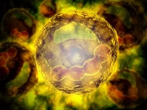
Budding yeast cell. Computer artwork of asection through a yeast ( Candida albicans )cell that is reproducing asexually. A daughtercell (top left) is budding from the parent cell

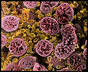
Background imageMitochondria Collection: Coloured SEM of mitochondria in ovarian cells

Coloured SEM of mitochondria in ovarian cells
Mitochondria. Coloured Scanning Electron Micro- graph (SEM) of mitochondria and smooth endoplasmic reticulum in an ovarian granulosa- lutein cell

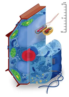
Background imageMitochondria Collection: Cell types, artwork

Cell types, artwork
Cell types. Cutaway artwork with a scale bar (upper right, in micrometres) showing the relative sizes of eukaryotic cells (those containing a nucleus) and prokaryotic cells (those lacking a nucleus)

Background imageMitochondria Collection: Plasma cells, TEM

Plasma cells, TEM
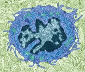
Plasma cells. Coloured transmission electron micrograph (TEM) of plasma cells. Plasma cells are mature B lymphocytes (white blood cells) that produce and secrete antibodies during an immune response

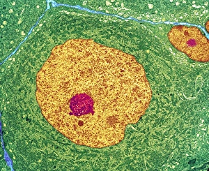
Background imageMitochondria Collection: Nerve cell, TEM

Nerve cell, TEM
Nerve cell. Coloured transmission electron micrograph (TEM) of a nerve cell body in cross- section. The cell has a large nucleus (yellow) and inner nucleolus (red)

Background imageMitochondria Collection: Mitochondrion, TEM

Mitochondrion, TEM
Mitochondrion. Coloured transmission electron micrograph (TEM) of mitochondria (green/blue) in heart muscle. Mitochondria are organelles found in the cytoplasm of eukaryotic cells

Background imageMitochondria Collection: Eye muscle, TEM C014 / 1468

Eye muscle, TEM C014 / 1468
Eye muscle. Transmission electron micrograph (TEM) of a section through a striated muscle cell from the ciliary muscle of a human eye

Background imageMitochondria Collection: Animal cell structure

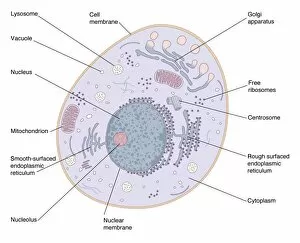
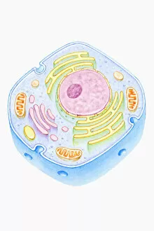
Animal cell structure. Artwork of a section through an animal cell. At lower centre is the nucleus, which contains the cells genetic information in the form of DNA (deoxyribonucleic acid)

Background imageMitochondria Collection: Microscopic view of animal cell

Microscopic view of animal cell

Background imageMitochondria Collection: TFAM transcription factor bound to DNA C015 / 7059

TFAM transcription factor bound to DNA C015 / 7059
TFAM transcription factor bound to DNA, molecular model. Human mitochondrial transcription factor A (TFAM, green) bound to a strand of DNA (deoxyribonucleic acid, blue and pink)

Background imageMitochondria Collection: Conceptual image of mitochondria

Conceptual image of mitochondria

Background imageMitochondria Collection: Muscle fibre structure, artwork

Muscle fibre structure, artwork
Muscle fibre. Computer artwork showing the structure of a muscle cell, also known as a muscle fibre. The cell is surrounded by a plasma membrane called the sarcolemma (cream)

Background imageMitochondria Collection: Hepatocyte liver cell, TEM

Hepatocyte liver cell, TEM
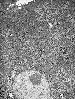
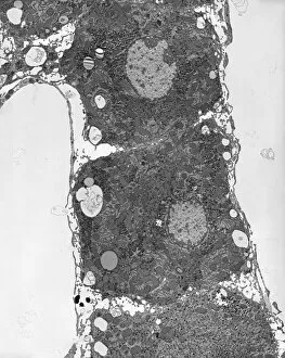
Hepatocyte liver cell. Transmission electron micrograph (TEM) of a section through an hepatocyte liver cell, showing rough and smooth endoplasmic reticulum (ER, dark lines)

Background imageMitochondria Collection: Liver tissue, TEM

Liver tissue, TEM
Liver tissue. Transmission electron micrograph (TEM) of a section through the liver, showing part of a radial cord of hepatocyte liver cells (dark) and the vascular sinusoids (white)

Background imageMitochondria Collection: Skeletal muscle, TEM C016 / 5369

Skeletal muscle, TEM C016 / 5369
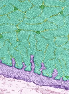
Skeletal muscle. Transmission electron micrograph (TEM) of a cross section through human skeletal (striated) muscle. Blocks of muscle (lighter grey) are surrounded by connective tissue (black)

Background imageMitochondria Collection: Skin cancer cell, SEM C016 / 4764

Skin cancer cell, SEM C016 / 4764
Skin cancer cell. Coloured ion-abrasion scanning electron micrograph (IA-SEM) of a melanoma cell. The large dark sphere is the cell nucleus, which contains the cells genetic information

Background imageMitochondria Collection: Mitochondria, TEM

Mitochondria, TEM
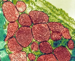
Mitochondria. Coloured transmission electron micrograph (TEM) of mitochondria (red) in heart muscle. Mitochondria are organelles found in the cytoplasm of eukaryotic cells

Background imageMitochondria Collection: Mitochondria, SEM

Mitochondria, SEM
Mitochondria. Coloured scanning electron micrograph (SEM) of mitochondria (red) in a kidney cell. Mitochondria are a type of organelle found in the cytoplasm of eukaryotic cells

Background imageMitochondria Collection: Cross section illustration of human cell

Cross section illustration of human cell

Background imageMitochondria Collection: Conceptual image of a plant cell and its components

Conceptual image of a plant cell and its components

Background imageMitochondria Collection: 3D structure of melanoma cell

3D structure of melanoma cell
3D structure of a melanoma cell derived by ion abrasion scanning electron microscopy

Background imageMitochondria Collection: Conceptual image of human cell

Conceptual image of human cell

Background imageMitochondria Collection: Human cell anatomy

Human cell anatomy

Background imageMitochondria Collection: Multiphoton fluorescence image of HeLa cells

Multiphoton fluorescence image of HeLa cells with cytoskeletal microtubules (magenta) and DNA (cyan)

Background imageMitochondria Collection: Microscopic view of Mitochondria

Microscopic view of Mitochondria. Mitochondria are the cells power producers. They convert energy into forms that are usable by the cell. Located in the cytoplasmcells activities

Background imageMitochondria Collection: Microscopic view of human cell

Microscopic view of human cell

Background imageMitochondria Collection: The interior of an eukaryotic cell

The interior of an eukaryotic cell. This is the most common generic type of cell and its present in all mammals. In the center is the nucleus (the perforated sphere)

Background imageMitochondria Collection: Conceptual image of the Golgi apparatus

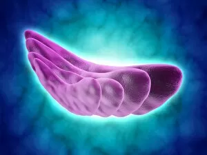
Conceptual image of the Golgi apparatus. The Golgi apparatus is an organelle found in most eukaryotic cells

Background imageMitochondria Collection: Comparative illustration of plant and animal cell anatomy (with labels)

Comparative illustration of plant and animal cell anatomy (with labels)

Background imageMitochondria Collection: Microscopic view of animal cell nucleus

Microscopic view of animal cell nucleus

Background imageMitochondria Collection: Illustration of cell

Illustration of cell
Medicine: Human anatomy, cell

Background imageMitochondria Collection: Model of animal cell, including cell nucleus, golgi body, lysosomes, centrioles, mitochondria

Model of animal cell, including cell nucleus, golgi body, lysosomes, centrioles, mitochondria, endoplasmic reticulum, ribosomes, cytoplasm, vesicles, thin plasma membrane

Background imageMitochondria Collection: Mitochondria, artwork

Mitochondria, artwork
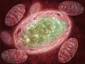
Mitochondria, computer artwork

Background imageMitochondria Collection: Intestinal gland cell, TEM C014 / 1441

Intestinal gland cell, TEM C014 / 1441
Intestinal gland cell. Transmission electron micrograph (TEM) of a section through an undifferentiated cell from the intestinal tract, showing extensive rough endoplasmic reticulum (ER)

Background imageMitochondria Collection: Intestinal gland cell, TEM C014 / 1440

Intestinal gland cell, TEM C014 / 1440
Intestinal gland cell. Transmission electron micrograph (TEM) of a section through an undifferentiated cell from the intestinal tract, showing extensive rough endoplasmic reticulum (ER)

Background imageMitochondria Collection: Plant cell, artwork F006 / 9876

Plant cell, artwork F006 / 9876
Plant cell, computer artwork

Background imageMitochondria Collection: P32 mitochondrial matrix protein F006 / 9454

P32 mitochondrial matrix protein F006 / 9454
P32 mitochondrial matrix protein, molecular model. Also known as SF2-associated p32 (SF2P32), this protein is found in the matrix of cellular mitochondria

Background imageMitochondria Collection: Mitochondrial structure, artwork F006 / 9207

Mitochondrial structure, artwork F006 / 9207
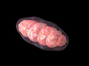
Mitochondrial structure. Computer artwork of a mitochondrion, showing a loop of mitochondrial DNA (deoxyribonucleic acid, mtDNA), the organelles genetic material

Background imageMitochondria Collection: Mitochondrial structure, artwork F006 / 9198

Mitochondrial structure, artwork F006 / 9198
Mitochondrial structure. Computer artwork of a mitochondrion, showing a loop of mitochondrial DNA (deoxyribonucleic acid, mtDNA), the organelles genetic material

Background imageMitochondria Collection: Cardiac muscle, light micrograph

Cardiac muscle, light micrograph
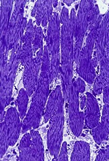
Cardiac muscle. Light micrograph of a section through muscle tissue from a heart, showing the cardiomyocyte cells. Heart muscle cells show central nuclei but the dominant feature is the abundance of

Background imageMitochondria Collection: Neuromuscular junction, artwork

Neuromuscular junction, artwork
Neuromuscular junction. Computer artwork showing the juntion between a neuron (nerve cell, light blue) and a muscle cell, known as a neuromuscular junction

Background imageMitochondria Collection: Lymphocyte white blood cell, TEM

Lymphocyte white blood cell, TEM
Lymphocyte white blood cell. Transmission electron micrograph (TEM) of a section through lymphocyte, showing the nucleus (large, round, centre), endoplasmic reticulum (ER)

Background imageMitochondria Collection: Steroid secreting cell, TEM

Steroid secreting cell, TEM
Steroid secreting cell. Transmission electron micrograph (TEM) of a section through a cell involved with steroid synthesis and secretion (steroidogenesis)

Background imageMitochondria Collection: Kidney mitochondria, TEM

Kidney mitochondria, TEM
Kidney mitochondria. Transmission electron micrograph (TEM) of a section through a kidney tubule, showing numerous mitochondria (blue)

Background imageMitochondria Collection: Eukaryote cell, artwork C016 / 6260

Eukaryote cell, artwork C016 / 6260
Eukaryote cell. Computer artwork showing the internal structure of a typical eukaryotic cell. Eukaryotes are organisms whose cells contain a membrane-bound nucleus (karyon, orange)

Background imageMitochondria Collection: VDAC-1 ion channel protein C015 / 8250

VDAC-1 ion channel protein C015 / 8250
VDAC-1 ion channel protein, molecular model. This is the human voltage-dependent anion-selective channel protein 1 (VDAC-1)

Background imageMitochondria Collection: Corpus luteum of ovary, light micrograph C016 / 0527

Corpus luteum of ovary, light micrograph C016 / 0527
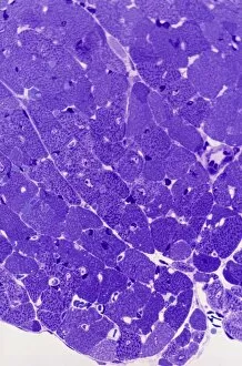
Corpus luteum of ovary. Light micrograph of a section through steroid-secreting luteal cells in the corpus luteum of an ovary. The corpus luteum develops from an ovulated follicle

Background imageMitochondria Collection: Heart muscle, light micrograph C016 / 0517

Heart muscle, light micrograph C016 / 0517
Heart muscle. Light micrograph of a section through heart (cardiac) muscle tissue, showing cardiomyocyte muscle cells. These cells have a centrally located nucleus and are branched


All Professionally Made to Order for Quick Shipping

Mitochondria, the powerhouse of the cell, play a vital role in various organisms. From budding yeast cells to ovarian cells, their presence is visually striking under scanning electron microscopy (SEM). The colored SEM image in ovarian cells showcases their intricate structure and diversity across different cell types. In nerve cells, transmission electron microscopy (TEM) reveals the fascinating architecture of mitochondria. These elongated organelles are strategically positioned along axons and dendrites, ensuring efficient energy supply for neuronal functions. The TEM images capture the complexity and importance within nerve cells. Moving beyond nerve cells, plasma cells also exhibit distinct mitochondrial features when observed through TEM. These specialized immune system components rely on robust energy production to carry out antibody synthesis efficiently. Their unique morphology highlights how mitochondria adapt to meet specific cellular demands. The eye muscle's TEM image further emphasizes the significance in highly active tissues. With an abundance of these organelles, eye muscles can sustain continuous contractions required for precise vision movements. Macrophage cells also showcase intriguing mitochondrial structures when examined using TEM techniques. As key players in our immune defense system, macrophages require substantial energy reserves to engulf pathogens effectively. Mitochondrial dynamics contribute significantly to this process by providing ample ATP generation. Under SEM imaging techniques specifically focused on mitochondria themselves, their distinctive shape becomes apparent—ranging from elongated tubules to spherical forms that vary across species and tissue types. Understanding animal cell structure necessitates studying not only individual organelles but also their interactions with other cellular components like DNA-binding proteins such as TFAM transcription factor bound to DNA shown in C015 / 7059 micrograph. Exploring various microscopic views allows us to appreciate the remarkable diversity and essential roles played by mitochondria across different cell types and organisms alike—a testament to their fundamental significance within biological systems.