Home > Science > SEM
Mitochondria, SEM

![]()
Wall Art and Photo Gifts from Science Photo Library
Mitochondria, SEM
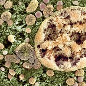
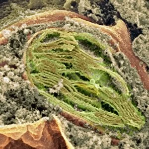
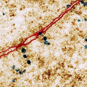
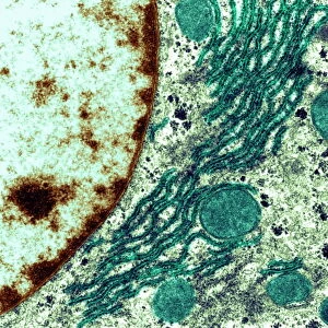
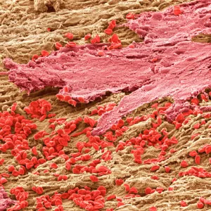
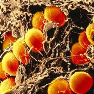
Mitochondria. Coloured scanning electron micrograph (SEM) of mitochondria (red) in a kidney cell. Mitochondria are a type of organelle found in the cytoplasm of eukaryotic cells. They oxidise sugars and fats to produce energy in a process called respiration. A mitochondrion has two membranes, a smooth outer membrane and a folded inner membrane. The folds of the inner membrane are called cristae, and it is here that the chemical reactions to produce energy take place. The orange structures seen here are the cell membranes of the kidney cells
Science Photo Library features Science and Medical images including photos and illustrations
Media ID 6303891
© DR DAVID FURNESS, KEELE UNIVERSITY/SCIENCE PHOTO LIBRARY
Cell Biology Cell Membrane Cross Section Cytology Energy Eukaryotic Folded Kidney Membrane Bound Membranes Membranous Metabolic Metabolism Mitochondria Mitochondrion Organelle Renal Respiration Structures Cells False Coloured Section Sectioned
EDITORS COMMENTS
This print showcases the intricate beauty of mitochondria, captured through a coloured scanning electron microscope (SEM). The image reveals a kidney cell, with the mitochondria highlighted in vibrant red. Mitochondria are essential organelles found within eukaryotic cells' cytoplasm, responsible for oxidising sugars and fats to generate energy in a process known as respiration. A mitochondrion consists of two membranes: an outer smooth membrane and an inner folded membrane. These folds, called cristae, play a crucial role in facilitating chemical reactions that produce energy. In this false-coloured image, the orange structures represent the cell membranes of the kidney cells surrounding these mighty powerhouses. The photograph not only provides us with a glimpse into cellular biology but also emphasizes the complexity and interconnectedness of life at its most fundamental level. It serves as a reminder that even within our own bodies, countless microscopic processes are constantly occurring to sustain our existence. Science Photo Library has once again captured nature's marvels with precision and artistry. This mesmerizing print is sure to captivate anyone interested in biology or those who appreciate the hidden wonders that lie beneath our skin.
MADE IN AUSTRALIA
Safe Shipping with 30 Day Money Back Guarantee
FREE PERSONALISATION*
We are proud to offer a range of customisation features including Personalised Captions, Color Filters and Picture Zoom Tools
SECURE PAYMENTS
We happily accept a wide range of payment options so you can pay for the things you need in the way that is most convenient for you
* Options may vary by product and licensing agreement. Zoomed Pictures can be adjusted in the Cart.