Fraud Blocker Skip to main content

Transmission Electron Micrograph Collection

"Exploring the Microscopic World: Unveiling Intricate Structures through Transmission Electron Micrograph" In the realm of science

Background imageTransmission Electron Micrograph Collection: Regenerating nerve cell, TEM

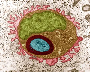
Regenerating nerve cell, TEM
Regenerating nerve cell. Coloured transmission electron micrograph (TEM) of a section through a nerve axon (blue) regenerating within a Schwann cell (light brown)

Background imageTransmission Electron Micrograph Collection: Fat cells, TEM

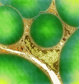
Fat cells, TEM
Fat cells. Coloured transmission electron micrograph (TEM) of a section through fat (adipose) tissue, consisting of fat-storing cells (adipocytes, green) supported by connective tissue

Background imageTransmission Electron Micrograph Collection: Regenerating nerve cell, TEM

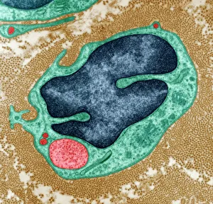
Regenerating nerve cell, TEM
Regenerating nerve cell. Coloured transmission electron micrograph (TEM) of a section through a nerve axon (yellow) regenerating within a Schwann cell (blue). The Schwann cells nucleus is black

Background imageTransmission Electron Micrograph Collection: E. coli bacterium, TEM

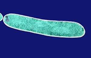
E. coli bacterium, TEM
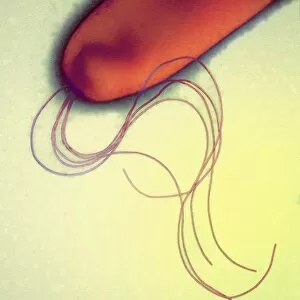
E. coli bacterium, coloured transmission electron micrograph (TEM). E. coli are Gram-negative rod-shaped bacteria that are part of the normal flora of the human gut

Background imageTransmission Electron Micrograph Collection: Plasma cells, TEM

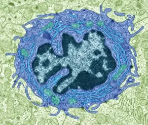
Plasma cells, TEM
Plasma cells. Coloured transmission electron micrograph (TEM) of plasma cells. Plasma cells are mature B lymphocytes (white blood cells) that produce and secrete antibodies during an immune response

Background imageTransmission Electron Micrograph Collection: Lung tissue, TEM

Lung tissue, TEM
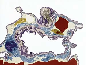
Lung tissue, coloured transmission electron micrograph (TEM). A single alveolus is at center. At top right is a pulmonary capillary containing a red blood cell (red) and a platelet (yellow)

Background imageTransmission Electron Micrograph Collection: Influenza virus particles, TEM

Influenza virus particles, TEM
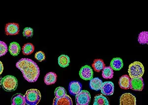
Influenza virus particles, coloured transmission electron micrograph (TEM). This virus is the cause of the respiratory disease influenza (flu) in humans and other animals

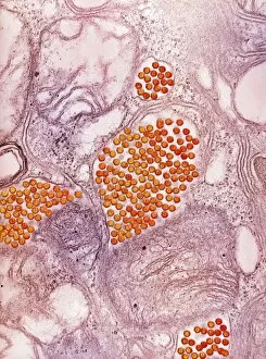
Background imageTransmission Electron Micrograph Collection: Hepatitis C viruses, TEM

Hepatitis C viruses, TEM
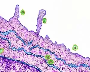
Hepatitis C viruses. Coloured transmission electron micrograph (TEM) of hepatitis C virus particles (green) infecting cultured liver cells (purple)

Background imageTransmission Electron Micrograph Collection: Eye muscle, TEM C014 / 1468

Eye muscle, TEM C014 / 1468
Eye muscle. Transmission electron micrograph (TEM) of a section through a striated muscle cell from the ciliary muscle of a human eye
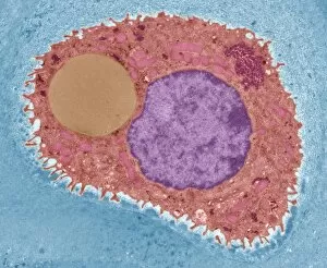
Background imageTransmission Electron Micrograph Collection: Purkinje nerve cell, TEM C014 / 0583

Purkinje nerve cell, TEM C014 / 0583
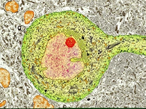
Purkinje nerve cell. Transmission electron micrograph (TEM) of a purkinje nerve cell (green) from the cerebellum of the brain, showing the cell body (centre) and its primary dendrite (cell process)

Background imageTransmission Electron Micrograph Collection: Intestinal microvilli, TEM

Intestinal microvilli, TEM
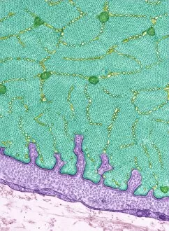
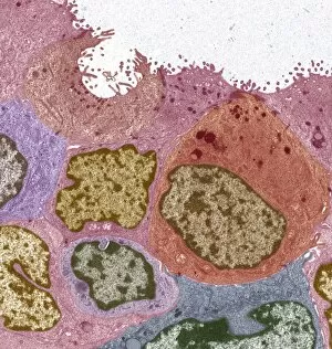
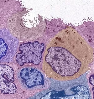
Intestinal microvilli, coloured transmission electron micrograph (TEM). The microvilli are finger-like projections (seen here in circular cross-section)

Background imageTransmission Electron Micrograph Collection: E. coli bacterium, TEM

E. coli bacterium, TEM
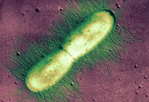
E. coli bacterium. Coloured transmission electron micrograph (TEM) of an Escherichia coli bacterium in the early stages of binary fission, the process by which the bacterium divides

Background imageTransmission Electron Micrograph Collection: HIV infected macrophage, SEM C018 / 8598

HIV infected macrophage, SEM C018 / 8598
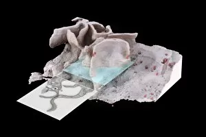
HIV infected macrophage. Coloured ion-abrasion scanning electron micrograph (IA-SEM) of a macrophage white blood cell infected with human immunodeficiency virus (HIV, red)

Background imageTransmission Electron Micrograph Collection: Pancreatic acinar cell

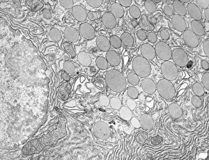
Pancreatic acinar cell. Transmission electron micrograph (TEM) of a section through an enzyme-secreting acinar cell in the human pancreas, showing part of the nucleus (round, far left)

Background imageTransmission Electron Micrograph Collection: Macrophage and lymphocytes, TEM

Macrophage and lymphocytes, TEM
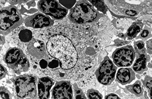
Macrophage and lymphocytes, transmission electron micrograph (TEM). Macrophage surrounded by lymphocytes in a lymph node. This view includes many intercellular contacts

Background imageTransmission Electron Micrograph Collection: Hepatocyte liver cell, TEM

Hepatocyte liver cell, TEM
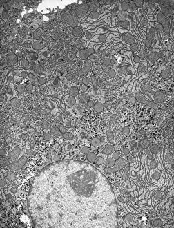
Hepatocyte liver cell. Transmission electron micrograph (TEM) of a section through an hepatocyte liver cell, showing rough and smooth endoplasmic reticulum (ER, dark lines)

Background imageTransmission Electron Micrograph Collection: Liver tissue, TEM

Liver tissue, TEM
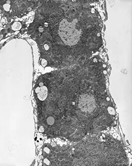
Liver tissue. Transmission electron micrograph (TEM) of a section through the liver, showing part of a radial cord of hepatocyte liver cells (dark) and the vascular sinusoids (white)

Background imageTransmission Electron Micrograph Collection: Cartilage cell, TEM C014 / 1433

Cartilage cell, TEM C014 / 1433
Cartilage cell. Transmission electron micrograph (TEM) of a section through a chondrocyte cell from hyaline cartilage of the trachea (windpipe)

Background imageTransmission Electron Micrograph Collection: Cartilage cell, TEM C014 / 1434

Cartilage cell, TEM C014 / 1434
Cartilage cell. Transmission electron micrograph (TEM) of a section through a chondrocyte cell from hyaline cartilage of the trachea (windpipe)

Background imageTransmission Electron Micrograph Collection: Influenza virus particles, TEM

Influenza virus particles, TEM
Influenza virus particles, coloured transmission electron micrograph (TEM). The virus consists of ribonucleic acid (RNA), surrounded by a nucleocapsid (blue) and a lipid envelope (green)

Background imageTransmission Electron Micrograph Collection: Golgi apparatus, TEM C017 / 8234

Golgi apparatus, TEM C017 / 8234
Golgi apparatus. Transmission electron micrograph (TEM) of Golgi apparatus from a Chlamydomonas sp. alga. Golgi are membrane-bound organelles that modify and package proteins

Background imageTransmission Electron Micrograph Collection: Skeletal muscle, TEM C016 / 5369

Skeletal muscle, TEM C016 / 5369
Skeletal muscle. Transmission electron micrograph (TEM) of a cross section through human skeletal (striated) muscle. Blocks of muscle (lighter grey) are surrounded by connective tissue (black)

Background imageTransmission Electron Micrograph Collection: Cardiac muscle, TEM

Cardiac muscle, TEM
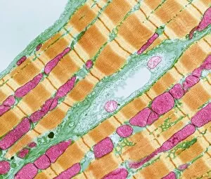
Cardiac muscle. Coloured transmission electron micrograph (TEM) of cardiac muscle fibrils (orange)from a healthy heart. Mitochondria (pink) supply the muscle cells with energy

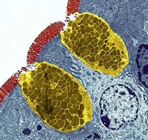
Background imageTransmission Electron Micrograph Collection: Goblet cells

Goblet cells. Coloured transmission electron micrograph (TEM) of a section through goblet cells in the lining of the small intestine, part of the digestive tract. They are full of mucus (yellow)

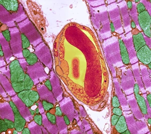
Background imageTransmission Electron Micrograph Collection: Cardiac muscle and capillary, TEM

Cardiac muscle and capillary, TEM
Cardiac muscle. Coloured transmission electron micrograph (TEM) of cardiac muscle fibrils (purple)from a healthy heart. Mitochondria (green) supply the muscle cells with energy

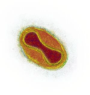
Background imageTransmission Electron Micrograph Collection: Smallpox virus particle, TEM

Smallpox virus particle, TEM
Smallpox virus particle. Coloured transmission electron micrograph (TEM) of a Variola major virus particle. Each particle consists of a DNA (deoxyribonucleic acid)

Background imageTransmission Electron Micrograph Collection: Infectious bronchitis virus (IBV), TEM

Infectious bronchitis virus (IBV), TEM
Infectious bronchitis virus (IBV) particles, coloured transmission electron micrograph (TEM). This virus is a member of the coronavirus family

Background imageTransmission Electron Micrograph Collection: Eastern equine encephalitis virus, TEM

Eastern equine encephalitis virus, TEM
Eastern equine encephalitis virus. Coloured transmission electron micrograph (TEM) of eastern equine encephalitis (EEE) virus particles (Alphavirus sp. orange) in the salivary gland of a mosquito

Background imageTransmission Electron Micrograph Collection: HIV particles, TEM

HIV particles, TEM
HIV (human immunodeficiency virus) particles, coloured transmission electron micrograph (TEM). Each particle consists of an RNA (ribonucleic acid, purple/brown) genome

Background imageTransmission Electron Micrograph Collection: H1N1 swine flu virus, TEM C018 / 8602

H1N1 swine flu virus, TEM C018 / 8602
H1N1 swine flu virus. Coloured transmission electron micrograph (TEM) of swine influenza (flu) virus particles (virions, yellow)

Background imageTransmission Electron Micrograph Collection: Blood clot, light micrograph

Blood clot, light micrograph
Blood clot. Light micrograph of a section through an arterial thrombus. Blood clots can form in an intact vessel that contains fatty deposits or one that is inflamed

Background imageTransmission Electron Micrograph Collection: Blood vessel, TEM

Blood vessel, TEM
Blood vessel. Coloured transmission electron micrograph (TEM) of a section through a blood vessel, showing platelets (blue) and red blood cells (red)

Background imageTransmission Electron Micrograph Collection: Eosinophil white blood cell, TEM C014 / 1438

Eosinophil white blood cell, TEM C014 / 1438
Eosinophil white blood cell. Transmission electron micrograph (TEM) of a section through an eosinophil. Eosinophils, like all white blood cells, are part of the bodys immune system

Background imageTransmission Electron Micrograph Collection: Bladder lining, TEM C014 / 1473

Bladder lining, TEM C014 / 1473
Bladder lining. Transmission electron micrograph (TEM) of a section through the epithelial cells that line of a human bladder, showing large flat vesicles (large, blue)

Background imageTransmission Electron Micrograph Collection: Intestinal gland cell, TEM C014 / 1441

Intestinal gland cell, TEM C014 / 1441
Intestinal gland cell. Transmission electron micrograph (TEM) of a section through an undifferentiated cell from the intestinal tract, showing extensive rough endoplasmic reticulum (ER)

Background imageTransmission Electron Micrograph Collection: Intestinal gland cell, TEM C014 / 1440

Intestinal gland cell, TEM C014 / 1440
Intestinal gland cell. Transmission electron micrograph (TEM) of a section through an undifferentiated cell from the intestinal tract, showing extensive rough endoplasmic reticulum (ER)

Background imageTransmission Electron Micrograph Collection: Cartilage cell, TEM C014 / 1432

Cartilage cell, TEM C014 / 1432
Cartilage cell. Transmission electron micrograph (TEM) of a section through a chondrocyte cell from hyaline cartilage of the trachea (windpipe)

Background imageTransmission Electron Micrograph Collection: Eosinophil white blood cell, TEM C014 / 1437

Eosinophil white blood cell, TEM C014 / 1437
Eosinophil white blood cell. Transmission electron micrograph (TEM) of a section through an eosinophil. Eosinophils, like all white blood cells, are part of the bodys immune system

Background imageTransmission Electron Micrograph Collection: Breast cancer, TEM F006 / 8633

Breast cancer, TEM F006 / 8633
Breast cancer. Coloured transmission electron micrograph (TEM) of a section through a ductal carcinoma (cancer) of the breast showing malignant epithelial cells

Background imageTransmission Electron Micrograph Collection: Breast cancer, TEM F006 / 8632

Breast cancer, TEM F006 / 8632
Breast cancer. Coloured transmission electron micrograph (TEM) of a section through a ductal carcinoma (cancer) of the breast showing malignant epithelial cells

Background imageTransmission Electron Micrograph Collection: Brain nerve cells, TEM C014 / 0357

Brain nerve cells, TEM C014 / 0357
Brain nerve cells. Transmission electron micrograph (TEM) of a section through brain tissue from the cerebral cortex, showing numerous neurons (nerve cells) surrounded by axons and dendrites

Background imageTransmission Electron Micrograph Collection: Eosinophil white blood cell, TEM C014 / 1439

Eosinophil white blood cell, TEM C014 / 1439
Eosinophil white blood cell. Transmission electron micrograph (TEM) of a section through an eosinophil. Eosinophils, like all white blood cells, are part of the bodys immune system

Background imageTransmission Electron Micrograph Collection: High-contrast direct DNA image, TEM

High-contrast direct DNA image, TEM
High-contrast direct DNA image. Coloured transmission electron micrograph (TEM) of the first high-contrast direct image of a bundle (fibre) of strands of DNA (deoxyribonucleic acid)

Background imageTransmission Electron Micrograph Collection: Reovirus particles, TEM C016 / 9443

Reovirus particles, TEM C016 / 9443
Reovirus particles. Coloured transmission electron micrograph (TEM) of numerous reovirus particles (virions, round). Reoviruses affect the gastrointestinal system and respiratory tract

Background imageTransmission Electron Micrograph Collection: Pseudomonas aeruginosa bacterium, SEM C016 / 9442

Pseudomonas aeruginosa bacterium, SEM C016 / 9442
Pseudomonas aeruginosa bacterium, coloured transmission electron micrograph (TEM). P. aeruginosa is a Gram-negative rod-shaped bacterium found in soil and water

Background imageTransmission Electron Micrograph Collection: Staphylococcus aureus bacteria, TEM C016 / 9458

Staphylococcus aureus bacteria, TEM C016 / 9458
Staphylococcus aureus bacteria. Coloured transmission electron micrograph (TEM) of a Staphylococcus aureus bacterium (round) in skin tissue. S

Background imageTransmission Electron Micrograph Collection: Yellow fever virus particles, TEM C016 / 9468

Yellow fever virus particles, TEM C016 / 9468
Yellow fever virus particles. Transmission electron micrograph (TEM) of yellow fever virus particles (virions, yellow). Yellow fever is endemic in tropical Africa and South America

Background imageTransmission Electron Micrograph Collection: Lassa virus particles, TEM C016 / 9409

Lassa virus particles, TEM C016 / 9409
Lassa virus particles. Transmission electron micrograph (TEM) of Lassa virus particles (virions, blue) amongst cell debris. This Arenavirus is the cause of Lassa fever


All Professionally Made to Order for Quick Shipping

"Exploring the Microscopic World: Unveiling Intricate Structures through Transmission Electron Micrograph" In the realm of science, transmission electron micrographs (TEM) have revolutionized our understanding of various biological entities. These powerful images provide a glimpse into the hidden intricacies that make up our world at an unimaginably small scale. One captivating TEM showcases regenerating nerve cells, offering hope for those seeking to understand and treat neurological disorders. The image captures the delicate process of nerve cell regrowth, highlighting their remarkable ability to heal and restore function. Another fascinating TEM reveals fat cells in all their glory - spherical structures filled with lipid droplets that play crucial roles in energy storage and insulation within our bodies. This microscopic view sheds light on how these adipocytes contribute to overall health and metabolism. Moving on, we encounter an E. coli bacterium captured by TEM, showcasing its distinctive rod-shaped structure. This notorious bacterium serves as both a model organism for research purposes and a cause of concern due to its potential pathogenicity. Delving deeper into the microscopic world, plasma cells come into focus through another mesmerizing TEM image. These specialized white blood cells produce antibodies vital for immune defense against invading pathogens - a testament to the intricate mechanisms at work within our bodies. The hauntingly beautiful transmission electron micrograph of influenza virus particles provides insight into one of humanity's greatest challenges - infectious diseases. Studying such images aids scientists in developing effective vaccines and antiviral treatments against these elusive viral foes. Hepatitis C viruses also reveal themselves under TEM scrutiny, reminding us of the ongoing battle against this chronic liver disease affecting millions worldwide. Understanding their structure helps researchers devise strategies for prevention and treatment. Shifting gears slightly from infections to anatomy, an eye muscle is showcased through TEM imagery – revealing its unique fiber arrangement responsible for precise eye movements essential for vision coordination. A Purkinje nerve cell takes center stage next; this neuron found in the cerebellum plays a crucial role in coordinating movement and balance.