
Flagella Collection
"Unveiling the Marvels of Flagella: A Journey into the Microscopic World" Embark on a captivating voyage through the intricate world of flagella


All Professionally Made to Order for Quick Shipping
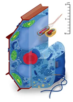
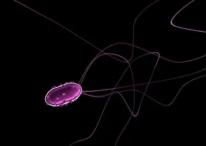
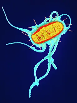
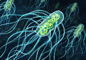
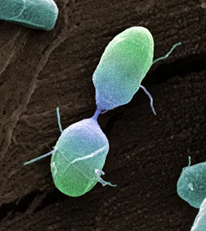
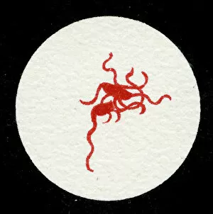
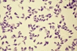
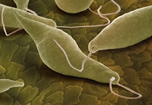
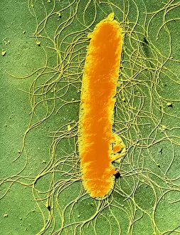
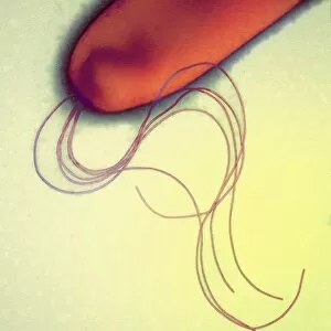
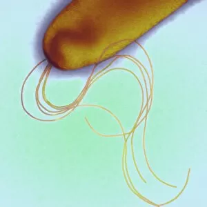
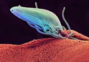
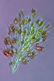
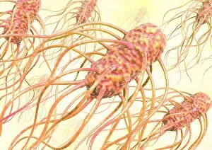
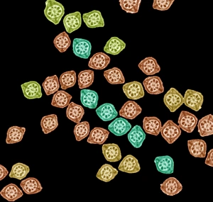
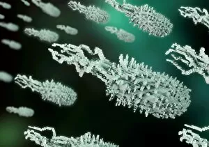
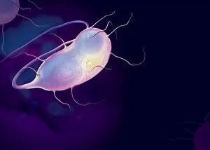
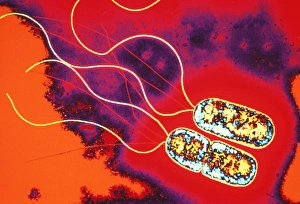
"Unveiling the Marvels of Flagella: A Journey into the Microscopic World" Embark on a captivating voyage through the intricate world of flagella, where cell types come alive in stunning artwork. Among these fascinating organisms are flagellate bacteria, such as the renowned E. Coli bacterium and Salmonella bacterium, captured dividing under the watchful eye of a scanning electron microscope (SEM). The SEM also reveals mesmerizing details of Salmonella typhimurium bacteria, showcasing their unique flagellar structures. Delving deeper into this microscopic realm, we encounter Escherichia coli bacteria depicted in vibrant colors through transmission electron microscopy (TEM), offering an artistic perspective on their beauty. Meanwhile, a lithograph from 1906 showcases a colony of Salmonella Typhi with Bacilli adorned by graceful flagella – a testament to early scientific exploration. Continuing our expedition, we encounter Euglena gracilis under SEM's lens; its elegant body propelled by whip-like flagella leaves us awestruck. Further along our journey lies Pyrococcus furiosus archaea - brought to life through breathtaking artwork - reminding us that even ancient microorganisms possess awe-inspiring adaptations like their own unique form of propulsion. Intriguingly diverse forms emerge as Giardia lamblia protozoa grace our vision in vivid detail via micrography. Their delicate yet powerful flagellar movements remind us that nature's wonders can be found even at microscopic scales. Finally, Proteus mirabilis bacterium captures our attention with its dynamic shape-shifting abilities and remarkable motility driven by multiple long and flexible flagella. Flagella serve as nature's propellers for these incredible organisms – enabling them to navigate their environments with precision and grace. As we conclude this extraordinary expedition into the world of flagella, we are left marveling at how these seemingly simple appendages hold immense significance in understanding cellular biology and the intricate workings of life itself.