
Backbone Collection (page 9)
"Unveiling the Backbone: From Dinosaurs to Humans" In this captivating journey through time and science, we explore the intricate marvel that is the backbone


All Professionally Made to Order for Quick Shipping
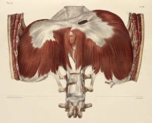
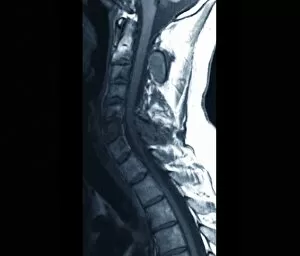
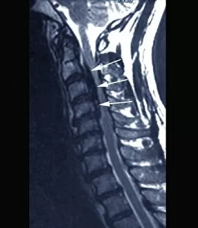
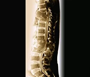
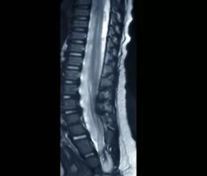
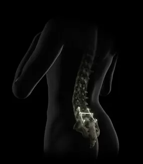
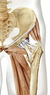
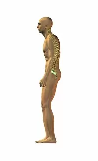
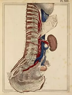
"Unveiling the Backbone: From Dinosaurs to Humans" In this captivating journey through time and science, we explore the intricate marvel that is the backbone. Starting with a diagram of the human spine in a side view, we witness the remarkable structure that supports our bodies and allows us to stand tall. Transporting us back millions of years, a 3D rendering of an Ankylosaurus dinosaur skeleton reminds us that even these ancient creatures possessed their own version of a backbone. The conceptual image of a human skull and spinal cord further emphasizes how this vital structure connects our brain to every part of our body. Continuing our exploration into prehistoric times, another 3D rendering reveals the awe-inspiring skeletal remains of a Tyrannosaurus Rex. Its massive backbone serves as evidence of its strength and dominance during its reign on Earth. Delving into history's scientific contributions, "The Science of Human Anatomy" by Bartholomeo Eustachi takes center stage. This groundbreaking work from centuries ago laid the foundation for understanding our bodies' inner workings. Shifting gears towards beauty and artistry, we encounter images showcasing bareback beauty – highlighting both vulnerability and strength embodied in one's posture. Leonardo Da Vinci's study on anatomy during the 15th century adds another layer to this multifaceted concept as medicine intertwines with artistic expression. Returning to modern times, detailed anatomical views present male skeletons from different perspectives – side view and perspective view – allowing us to appreciate how each bone fits together like puzzle pieces forming an unyielding support system for life itself. As we conclude this visual expedition through time and knowledge, it becomes evident that whether it be dinosaurs or humans, past or present; there is something truly extraordinary about the backbone - an enduring symbol representing resilience, adaptability, and evolution across species throughout history.