Home > Popular Themes > Human Body
Hip bones, muscles and ligaments, artwork C016 / 7018

![]()
Wall Art and Photo Gifts from Science Photo Library
Hip bones, muscles and ligaments, artwork C016 / 7018
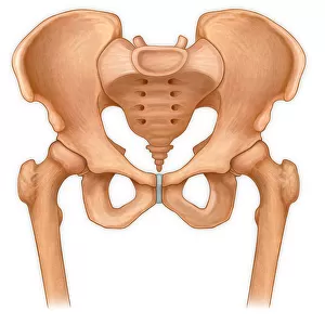
Hip bones and thigh muscles. Artwork of a frontal (anterior) view of the left hip, showing bones and some thigh muscles (red). At lower right is the femur (thigh bone). The head of the femur (ball-shaped) is covered in cartilage (grey) that cushions it in the acetabulum, the hip socket in the pelvis. The pelvis bones that form part of the hip joint are the ischium and pubis (centre left) and the ilium (upper centre). At top left are bones of the lower spine (coccyx, sacrum, lumbar vertebrae). The muscles shown are attached to the tuberosity of the ischium. These include the adductor magnus and the hamstring muscles (biceps femoris, semimembranosus, semitendinosus)
Science Photo Library features Science and Medical images including photos and illustrations
Media ID 9244287
© D & L GRAPHICS / SCIENCE PHOTO LIBRARY
Acetabulum Adductor Magnus Anterior Articulation Backbone Biceps Femoris Bones Cartilage Coccyx Femoral Femoral Head Femur Frontal Gluteus Minimus Hamstrings Hip Socket Ilium Ischium Joint Ligament Ligaments Lumbar Muscles Pelvic Pubis Sacrum Semimembranosus Semitendinosus Tail Bone Tailbone Thigh Bone Inguinal Ligament Obturator Externus Pelvis Vertebrae
EDITORS COMMENTS
This artwork, titled "Hip bones, muscles and ligaments" offers a detailed frontal view of the left hip, showcasing its intricate anatomy. Against a pristine white background, this illustration provides an invaluable glimpse into the complexity of our musculoskeletal system. At the forefront of this composition are the hip bones and thigh muscles depicted in vibrant red hues. The femur, or thigh bone, takes center stage at the lower right corner. Its ball-shaped head is covered in grey cartilage that acts as a cushion within the acetabulum—the socket where it articulates with the pelvis. Speaking of which, we can observe three main pelvic bones forming part of this joint: the ischium and pubis towards the center-left and upper-center respectively, along with the ilium. Meanwhile, at top left lie various vertebrae from our lower spine—coccyx, sacrum, and lumbar vertebrae—highlighting their connection to hip function. The artist has also thoughtfully included several key muscles attached to the tuberosity of the ischium. These include notable ones like adductor magnus and hamstring muscles such as biceps femoris, semimembranosus, and semitendinosus. Overall, this visually striking print encapsulates both beauty and scientific precision—a testament to human anatomy's marvels while serving as an educational resource for those seeking a deeper understanding of our remarkable bodies.
MADE IN AUSTRALIA
Safe Shipping with 30 Day Money Back Guarantee
FREE PERSONALISATION*
We are proud to offer a range of customisation features including Personalised Captions, Color Filters and Picture Zoom Tools
SECURE PAYMENTS
We happily accept a wide range of payment options so you can pay for the things you need in the way that is most convenient for you
* Options may vary by product and licensing agreement. Zoomed Pictures can be adjusted in the Cart.