Fraud Blocker Skip to main content

Pubis Collection

The pubis, a crucial part of the human anatomy, has been depicted and studied throughout history

Background imagePubis Collection: Venus of Willendorf

Venus of Willendorf
6360261 Venus of Willendorf by Prehistoric; Naturhistorisches Museum, Vienna, Austria; (add.info.: Venus of Willendorf, 110 mm.: woman statue made in limestone and painted with ochre)

Background imagePubis Collection: Picture No. 10873599

Picture No. 10873599
Human Crab Louse (Phthirus pubis) Date:

Background imagePubis Collection: Buttock muscles, artwork C013 / 4414

Buttock muscles, artwork C013 / 4414
Buttock muscles. Computer artwork of a posterior view of the hips showing some of the muscles (red) of the buttocks. These are shown attached to the femurs (thigh bones) and the pelvis bones

Background imagePubis Collection: Lumbar region anatomy engraving 1866

Lumbar region anatomy engraving 1866
Atlas d anatomie descriptive du corps humain C. Bonamy - Paul Broca Victor Masson et Fils Paris 1866

Background imagePubis Collection: Cross section biomedical illustration of female type pelvis

Cross section biomedical illustration of female type pelvis

Background imagePubis Collection: Picture No. 10873600

Picture No. 10873600
Human Crab Louse (Phthirus pubis) Date:

Background imagePubis Collection: Hip bones, artwork C016 / 7015

Hip bones, artwork C016 / 7015
Hip bones. Artwork of a frontal (anterior) view of the left hip, showing the bones that articulate to form this joint. At lower right is the femur (thigh bone)

Background imagePubis Collection: Hip bones and thigh muscles, artwork C016 / 7016

Hip bones and thigh muscles, artwork C016 / 7016
Hip bones and thigh muscles, artwork

Background imagePubis Collection: Hip bones, muscles and ligaments, artwork C016 / 7018

Hip bones, muscles and ligaments, artwork C016 / 7018
Hip bones and thigh muscles. Artwork of a frontal (anterior) view of the left hip, showing bones and some thigh muscles (red). At lower right is the femur (thigh bone)

Background imagePubis Collection: Bones of the torso, artwork

Bones of the torso, artwork
Bones of the torso and neck, computer artwork. At centre is the spine. The cervical (neck) spine is yellow, the thoracic (upper) spine is red and the lumbar (lower) spine is maroon

Background imagePubis Collection: Fractured hip, artwork

Fractured hip, artwork
Fractured hip. Computer artwork of a fracture in the head of the femur (thigh bone)

Background imagePubis Collection: Pelvic floor, artwork

Pelvic floor, artwork
Male pelvic floor. Computer artwork showing the levator ani muscle of the pelvic floor. The pelvic floor provides support to the pelvic organs, such as the bladder and prostate

Background imagePubis Collection: Lower body anatomy, artwork

Lower body anatomy, artwork
Lower body anatomy. Computer artwork showing the bones (cream), muscles (pink), nerves (yellow) and ligaments and tendons (white) of the lower body

Background imagePubis Collection: Hip ligaments, artwork C013 / 4434

Hip ligaments, artwork C013 / 4434
Hip ligaments. Computer artwork of a posterior oblique view of the hip bones and ligaments (white). At upper right the lumbar spine ends in the sacrum and coccyx (tailbone)

Background imagePubis Collection: Hip ligaments, artwork C013 / 4433

Hip ligaments, artwork C013 / 4433
Hip ligaments. Computer artwork of a posterior view of the hip bones and ligaments (white). At centre the lumbar spine ends in the sacrum and coccyx (tailbone)

Background imagePubis Collection: Hip ligaments, artwork C013 / 4432

Hip ligaments, artwork C013 / 4432
Hip ligaments. Computer artwork of an anterior view of the hip bones and ligaments (white). At centre the lumbar spine ends in the sacrum and coccyx (tailbone)

Background imagePubis Collection: Hip anatomy, artwork C013 / 4430

Hip anatomy, artwork C013 / 4430
Hip anatomy. Computer artwork of the head of the left femur (centre right) articulating with the left-hand side of the pelvis to form the hip joint

Background imagePubis Collection: Leg muscles, artwork C013 / 4429

Leg muscles, artwork C013 / 4429
Leg muscles. Computer artwork of the bones and outer muscles of the left leg, seen from the front (anterior view) at an oblique angle looking downwards

Background imagePubis Collection: Thigh hamstring muscles, artwork C013 / 4428

Thigh hamstring muscles, artwork C013 / 4428
Thigh hamstring muscles. Computer artwork of a posterior view of two of the three hamstring muscles (red) of the thighs. The muscles are the semimembranosus (inner) and semitendinosus (outer)

Background imagePubis Collection: Iliopsoas muscles, artwork C013 / 4425

Iliopsoas muscles, artwork C013 / 4425
Iliopsoas muscles. Computer artwork of an anterior (frontal) view of the hips, showing the inner hip muscles (red), the ilioposas

Background imagePubis Collection: Buttock muscles, artwork C013 / 4423

Buttock muscles, artwork C013 / 4423
Buttock muscles. Computer artwork of a posterior oblique view of the hips, showing some of the muscles (red) of the buttocks

Background imagePubis Collection: Buttock muscles, artwork C013 / 4424

Buttock muscles, artwork C013 / 4424
Buttock muscles. Computer artwork of a posterior view of the hips showing some of the muscles (red) of the buttocks. These are shown attached to the femurs (thigh bones) and the pelvis bones

Background imagePubis Collection: Buttock muscles, artwork C013 / 4422

Buttock muscles, artwork C013 / 4422
Buttock muscles. Computer artwork of a posterior oblique view of the hips showing some of the muscles (red) of the buttocks. These are shown attached to the femurs (thigh bones) and the pelvis bones

Background imagePubis Collection: Buttock muscles, artwork C013 / 4420

Buttock muscles, artwork C013 / 4420
Buttock muscles. Computer artwork of a posterior view of the hips showing some of the muscles (red) of the buttocks. These are shown attached to the femurs (thigh bones) and the pelvis bones

Background imagePubis Collection: Buttock muscles, artwork C013 / 4421

Buttock muscles, artwork C013 / 4421
Buttock muscles. Computer artwork of a side view (front of body at left) of the hips showing some of the muscles (red) of the buttocks

Background imagePubis Collection: Buttock muscles, artwork C013 / 4419

Buttock muscles, artwork C013 / 4419
Buttock muscles. Computer artwork of a posterior oblique view of the hips showing some of the muscles (red) of the buttocks. These are shown attached to the femurs (thigh bones) and the pelvis bones

Background imagePubis Collection: Buttock muscles, artwork C013 / 4415

Buttock muscles, artwork C013 / 4415
Buttock muscles. Computer artwork of a posterior oblique view of the hips showing some of the muscles (red) of the buttocks. These are shown attached to the femurs (thigh bones) and the pelvis bones

Background imagePubis Collection: Buttock muscles, artwork C013 / 4416

Buttock muscles, artwork C013 / 4416
Buttock muscles. Computer artwork of a posterior oblique view of the hips showing some of the muscles (red) of the buttocks. These are shown attached to the femurs (thigh bones) and the pelvis bones

Background imagePubis Collection: Coloured X-ray of hip fracture due to osteoporosis

Coloured X-ray of hip fracture due to osteoporosis
Osteoporosis fracture. Coloured X-ray of the hip of an elderly woman with a fracture (centre left, yellow) caused by osteoporosis (brittle bone disease)

Background imagePubis Collection: Hip fracture due to osteoporosis

Hip fracture due to osteoporosis
Osteoporosis fracture. Coloured X-ray of the hip of an elderly woman with a fracture (centre left, orange) caused by osteoporosis (brittle bone disease)


All Professionally Made to Order for Quick Shipping

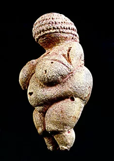
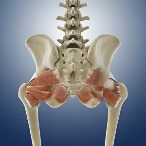
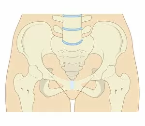
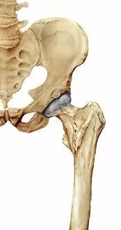
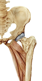
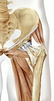
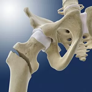
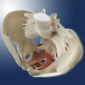
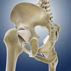
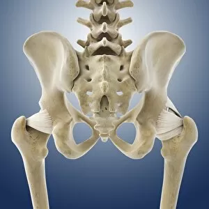
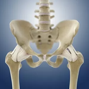
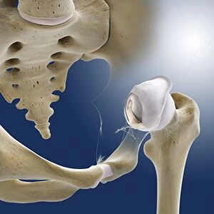
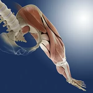
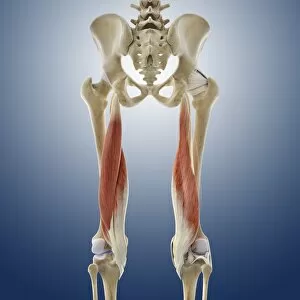
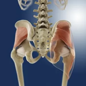
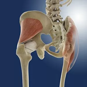
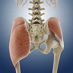
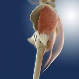
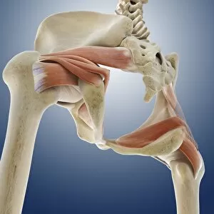
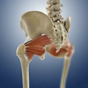
The pubis, a crucial part of the human anatomy, has been depicted and studied throughout history. One notable representation is found in the Venus of Willendorf, an ancient figurine showcasing exaggerated buttock muscles and a prominent pubic area (Picture No. 10873599). This small sculpture serves as a testament to the significance placed on fertility and sexuality in early civilizations. Delving deeper into its anatomical context, we find engravings from 1866 that illustrate the lumbar region's connection to the pubis (Lumbar region anatomy engraving 1866). Biomedical illustrations provide cross-sections of female pelvises, shedding light on various conditions such as urinary incontinence (Cross section biomedical illustration of urinary incontinence in female) or normal bladder function (Cross section biomedical illustration of normal bladder in female). Exploring further within this realm, artwork dedicated to understanding pelvic anatomy showcases intricate details about hip bones and thigh muscles (Hip bones and thigh muscles, artwork C016 / 7015). These depictions also highlight how ligaments interact with these structures for stability and movement purposes (Hip bones, muscles and ligaments, artwork C016 / 7018). In essence, whether it be through ancient sculptures like the Venus of Willendorf or modern medical illustrations depicting pelvic anatomy intricacies (Female pelvic anatomy), studying the pubis allows us to unravel both artistic expressions and scientific knowledge. It reminds us that our bodies are marvelously complex systems deserving exploration from multiple perspectives.