Home > Popular Themes > Human Body
Buttock muscles, artwork C013 / 4420

![]()
Wall Art and Photo Gifts from Science Photo Library
Buttock muscles, artwork C013 / 4420
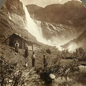
Buttock muscles. Computer artwork of a posterior view of the hips showing some of the muscles (red) of the buttocks. These are shown attached to the femurs (thigh bones) and the pelvis bones. At lower left is the largest muscle, the gluteus maximus, one of the strongest muscles in the body. Also seen at upper left, above the gluteus maximus, is the gluteus medius. At centre right (the right hip) only the smaller gluteus medius muscle is shown. The lower part of the spine and its vertebrae is at upper centre, ending in the sacrum and coccyx. Some of the lower ribs are also seen at top right and left
Science Photo Library features Science and Medical images including photos and illustrations
Media ID 9195969
© SPRINGER MEDIZIN/SCIENCE PHOTO LIBRARY
Backbone Bones Buttocks Coccyx Connective Tissue Coxal Bone Femoral Femur Femurs From Behind Gluteal Muscles Gluteals Gluteus Maximus Gluteus Medius Hip Bone Hips Iliac Crest Ilium Innominate Bone Ischium Joint Joints Legs Ligament Ligaments Lower Back Lower Limbs Lumbar Vertebrae Muscles Muscular System Musculoskeletal System Pelvic Posterior Pubis Sacrum Skeletal Tailbone Tendon Tendons Thigh Thigh Bone Upper Leg Vertebral Column Musculature Pelvis
EDITORS COMMENTS
This print showcases the intricate network of muscles in the buttocks, providing a detailed view of their attachment to the femurs and pelvis bones. The vibrant red hues highlight the strength and power of these muscles, particularly emphasizing the largest one known as gluteus maximus. Renowned for its robustness, this muscle is considered one of the strongest in our entire body. The composition also reveals another significant muscle called gluteus medius, positioned above the gluteus maximus on the left side. However, on the right hip, only a smaller portion of this muscle is visible. Alongside these muscular structures lies an array of skeletal elements including lower ribs at both top corners and parts of the vertebral column extending upwards towards upper center. This male skeleton illustration not only delves into biological intricacies but also offers insights into human anatomy from behind. It explores various components such as connective tissues, tendons, ligaments, and joints that contribute to a healthy musculoskeletal system. Additionally, it highlights key features like pubis bone, sacrum and coccyx (tailbone), lumbar vertebrae (lower back), ilium (part of pelvis), ischium (hip bone), and iliac crest. Overall, this artwork provides an educational glimpse into our body's complex structure while celebrating its remarkable capabilities through vivid imagery captured by Springer Medizin/Science Photo Library.
MADE IN AUSTRALIA
Safe Shipping with 30 Day Money Back Guarantee
FREE PERSONALISATION*
We are proud to offer a range of customisation features including Personalised Captions, Color Filters and Picture Zoom Tools
SECURE PAYMENTS
We happily accept a wide range of payment options so you can pay for the things you need in the way that is most convenient for you
* Options may vary by product and licensing agreement. Zoomed Pictures can be adjusted in the Cart.