
Femurs Collection
"Femurs: The Strong Foundation of the Hip Bones" The femur, also known as the thigh bone, plays a vital role in our skeletal structure


All Professionally Made to Order for Quick Shipping
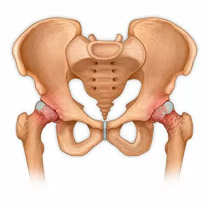
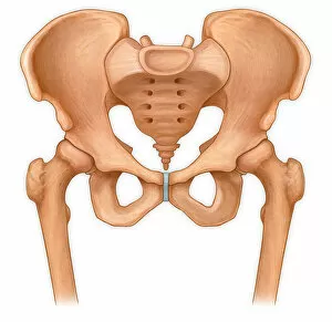
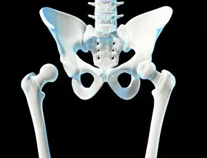
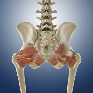
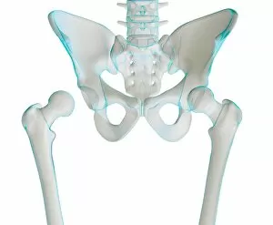
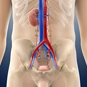
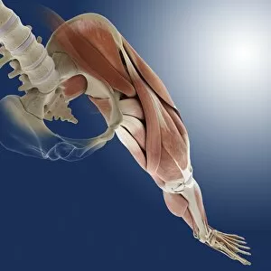
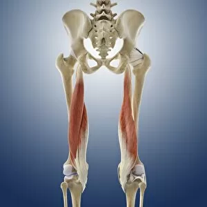
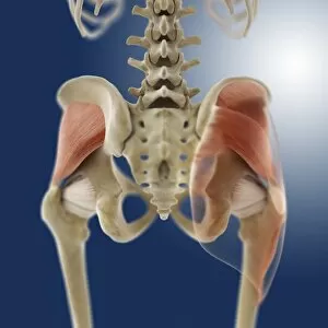
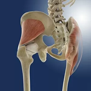
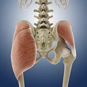
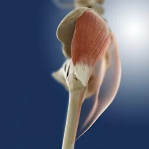
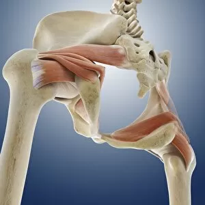
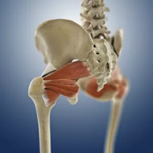
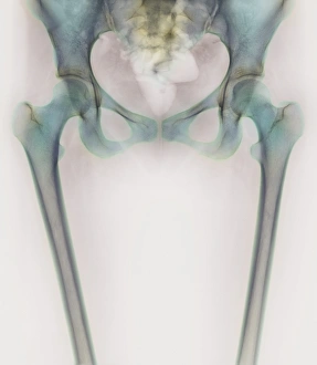
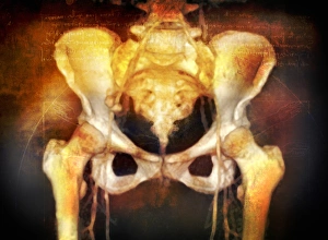
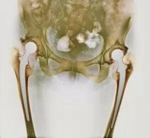
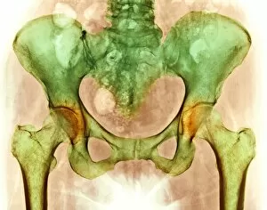
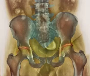
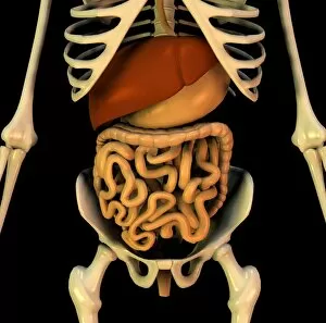
"Femurs: The Strong Foundation of the Hip Bones" The femur, also known as the thigh bone, plays a vital role in our skeletal structure. As we delve into its significance, let's explore various aspects that make it truly remarkable. In an X-ray image capturing the normal anterior view of the pelvis with hip bones, one can witness the robustness and intricate design of this essential bone. Its sheer strength provides stability to our lower body and facilitates smooth movement. However, not all they can free from ailments. An anterior view showcasing arthritis and osteophytes on femoral heads reminds us of how crucial it is to take care of these precious joints. This visual representation serves as a reminder to prioritize joint health through proper exercise and nutrition. Artwork depicting hip joint bones and anatomy further emphasizes the complexity involved in this region. With artwork C014 / 2032 beautifully illustrating this intricate network, we gain a deeper understanding of how everything fits together seamlessly. Moving beyond just bones, buttock muscles (artwork C013 / 4414) play a significant role in supporting our hips during movement. These powerful muscles work hand-in-hand with our femurs to provide stability while walking or running. When exploring the lower half of the human skeleton from a front view (jaw, spinal column, rib cage, pelvis), it becomes evident that without strong femurs (alongside arms and hands), maintaining balance would be nearly impossible. Our legs owe their strength to these mighty bones. Returning once again to artwork C014 / 2031 showcasing hip joint anatomy allows us to appreciate its complexity even more deeply. Understanding its intricacies helps medical professionals diagnose issues accurately for effective treatment plans. Shifting gears slightly towards internal organs' illustration - single-kidney anatomy depicted by artwork C016 / 2901 & C016 / 2900 - highlights how close these vital organs lie next to our sturdy femurs.