
Femoral Collection
"Femoral: Exploring the Intricacies of the Nervous System and Hip Replacement" Delving into the fascinating world anatomy


All Professionally Made to Order for Quick Shipping
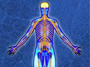
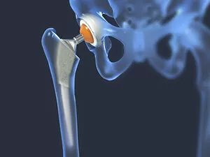
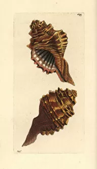
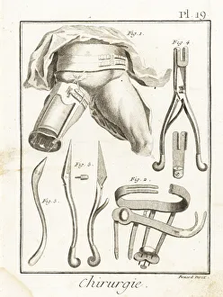
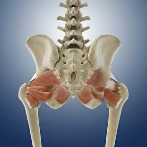
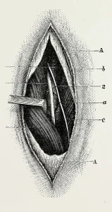
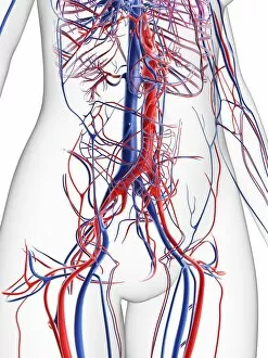
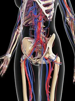
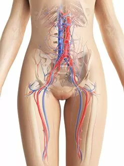
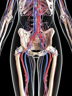
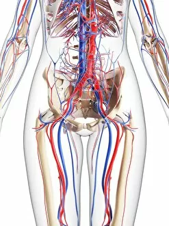
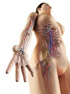
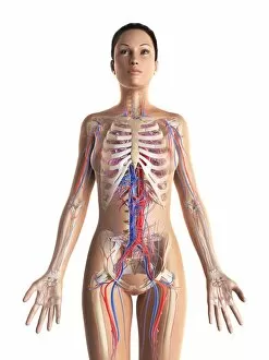
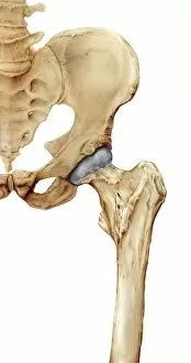
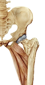
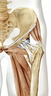
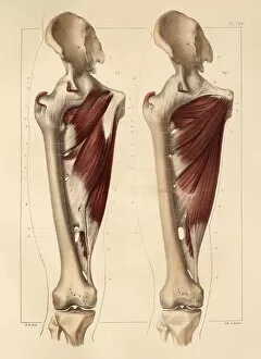
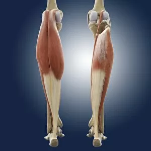
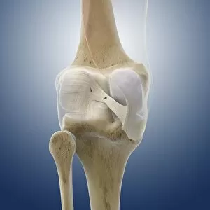
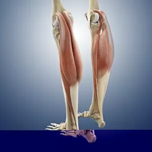
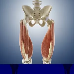
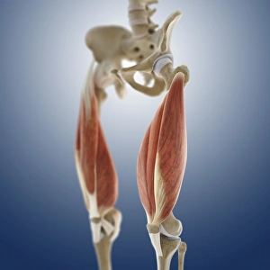
"Femoral: Exploring the Intricacies of the Nervous System and Hip Replacement" Delving into the fascinating world anatomy, we uncover its crucial role in our nervous system. A visual representation of hip replacement showcases the intricate surgical procedure that can restore mobility and alleviate pain. The mesmerizing Cymatium femorale shell reminds us of nature's incredible design, mirroring the structure of our own femur bone. Jean Louis Petit's groundbreaking machine to compress highlights his innovative approach to treating femoral fractures in the 18th century. Step back in time with an artistic illustration depicting pelvic-femoral muscles from a 1831 artwork, showcasing their complexity and importance. Discovering the strength behind our movements, artwork C013/4414 beautifully captures buttock muscles' role in supporting the femoral region. An engraving from 1866 provides a detailed glimpse into knee region anatomy, shedding light on how it connects with the intricate network surrounding it. Unveiling another layer of knowledge, an engraving from 1866 explores femoral region anatomy, unraveling its connection to various bodily functions. Dive deeper into understanding pelvic-femoral region anatomy through an enchanting engraving from 1866 that reveals its interconnectedness within our body systems. Embark on a journey through time as we explore pelvic-femoral region anatomy once again with another captivating engraving from 1866 - marvel at how far medical illustrations have come. In summary, "Femoral" encompasses various aspects such as nervous system involvement, hip replacement procedures illustrated visually or historically (Jean Louis Petits machine), intriguing natural references (Cymatium Femorale), depictions of muscle groups like pelvic-femoral and buttock muscles (artwork C013/4414), along with engravings from 1866 that shed light on knee and femoral region anatomy