
Bone Marrow Collection
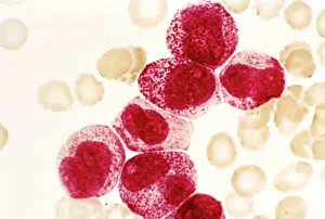
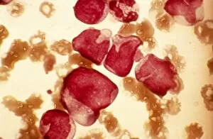
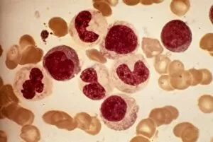
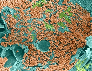
"Unveiling the Mysteries of Bone Marrow: From Ancient Egypt to Modern Medicine" Acute promyelocytic leukaemia, a rare form of blood cancer


All Professionally Made to Order for Quick Shipping
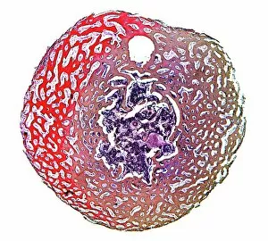
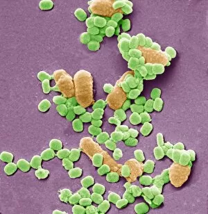
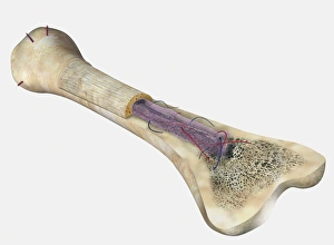
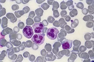
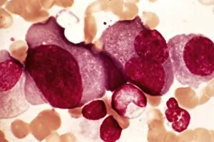
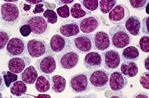
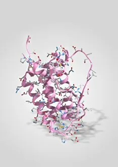
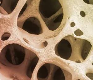
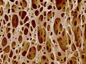
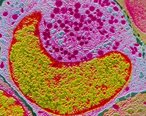
"Unveiling the Mysteries of Bone Marrow: From Ancient Egypt to Modern Medicine" Acute promyelocytic leukaemia, a rare form of blood cancer, has researchers turning their attention towards bone marrow for potential treatments. (Micrograph) Delving into the depths of spongy bone, a light micrograph reveals the intricate network that houses precious bone marrow. Journey back in time to ancient Egypt's Valley of the Queens, where Queen Nefertari's tomb holds secrets about the significance in their beliefs and rituals. (Luxor) Exploring under an electron microscope, we discover virus particles and bacteria lurking within bone marrow - shedding light on its role as a battleground against infections. A cross-section diagram showcases the complexity of human bone marrow within our femur - highlighting osteons, veins, and vital hematopoietic cells responsible for blood production. Witnessing cultural practices in Kenya's Samburu tribe, a young boy draws sustenance by sucking nutritious marrow straight from a cow's leg bone. Unraveling the anatomy of human bone marrow unveils its crucial function as a source for stem cells involved in tissue repair and regeneration. In culinary traditions worldwide, grilled bones are utilized to create rich brown stock - capturing both flavor and nutrients from deep within the marrow-filled cavities. Peering through another lens with a light micrograph brings forth stunning details showcasing the beauty hidden within our own bones' vibrant inner world - filled with life-sustaining marrow. From medical breakthroughs to cultural customs and gastronomic delights, exploring "bone marrow" opens doors to fascinating realms that intertwine science, history, health benefits, and diverse human experiences across time and continents.