Humeral bone marrow cavity, artwork C016 / 2888

![]()
Wall Art and Photo Gifts from Science Photo Library
Humeral bone marrow cavity, artwork C016 / 2888
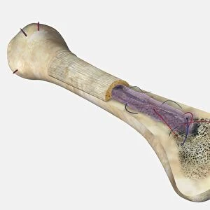
Humeral bone marrow cavity. Artwork of a sectioned upper arm bone (humerus), one of the long bones of the skeleton. These carry most of the load in the arms and legs. The outer layer is compact bone (cortical bone), with an interior of cancellous bone (spongy bone). The interior of long bones includes the medullary cavity (blood vessel shown) where bone marrow is located. The medullary cavity of long bones contains yellow marrow, with red marrow in each rounded end of the bone (the epiphysis, top). The epiphyseal line lies between the epiphysis and the shaft (diaphysis)
Science Photo Library features Science and Medical images including photos and illustrations
Media ID 9202717
© SPRINGER MEDIZIN/SCIENCE PHOTO LIBRARY
Arterial Bone Marrow Cancellous Bone Cavity Compact Bone Cortical Bone Cutaway Diaphysis Epiphysis Humerus Inside Interior Layer Layers Long Bone Osteology Shaft Skeletal Spongy Bone Tissue Artery Blood Supply Blood Vessel Blue Background Circulation Circulatory System Medullary Cavity Osseous Tissue Section Sectioned
EDITORS COMMENTS
This print showcases the intricate details of a humeral bone marrow cavity, providing a fascinating glimpse into the inner workings of our skeletal system. The artwork, labeled C016 / 2888, depicts a sectioned upper arm bone known as the humerus - one of the long bones responsible for bearing most of the load in our arms and legs. The outer layer of this bone is composed of compact bone or cortical bone, which provides strength and protection. Beneath this protective layer lies cancellous or spongy bone, with its characteristic porous structure that aids in shock absorption. Within the medullary cavity depicted in this image resides the vital component known as bone marrow. This hollow space serves as a home to yellow marrow while red marrow is concentrated within each rounded end called epiphysis. The epiphyseal line demarcates these ends from the shaft or diaphysis. Highlighted against a serene blue background, blood vessels are prominently displayed throughout this artwork, showcasing their crucial role in supplying nutrients and oxygen to support healthy tissue function. This stunning illustration not only offers an insight into human anatomy but also emphasizes the significance of circulation and blood production within our bodies. It serves as a reminder that beneath our skin lies an intricately designed framework that enables us to move and thrive every day.
MADE IN AUSTRALIA
Safe Shipping with 30 Day Money Back Guarantee
FREE PERSONALISATION*
We are proud to offer a range of customisation features including Personalised Captions, Color Filters and Picture Zoom Tools
SECURE PAYMENTS
We happily accept a wide range of payment options so you can pay for the things you need in the way that is most convenient for you
* Options may vary by product and licensing agreement. Zoomed Pictures can be adjusted in the Cart.