Fraud Blocker Skip to main content

Bread Mould Collection

"Bread Mould: Unveiling the Intricate World of Fungal Growth" Delving into the microscopic realm

Background imageBread Mould Collection: Mold for a Eulogia (Blessing) Bread, 600s-900s (wood)

Mold for a Eulogia (Blessing) Bread, 600s-900s (wood)
491396 Mold for a Eulogia (Blessing) Bread, 600s-900s (wood) by Byzantine School; h:8.80 d:1.90 cm; Cleveland Museum of Art, OH

Background imageBread Mould Collection: Bread mould, SEM C017 / 7127

Bread mould, SEM C017 / 7127
Bread mould. Coloured scanning electron micrograph (SEM) of a sporangium of a bread mould (Mucor sp.). Sporangia are asexual reproductive structures where the moulds spores develop

Background imageBread Mould Collection: Bread mould, SEM C017 / 7129

Bread mould, SEM C017 / 7129
Bread mould. Coloured scanning electron micrograph (SEM) of a sporangium of a bread mould (Mucor sp.). Sporangia are asexual reproductive structures where the moulds spores develop

Background imageBread Mould Collection: Bread mould, SEM C017 / 7128

Bread mould, SEM C017 / 7128
Bread mould. Coloured scanning electron micrograph (SEM) of a sporangium of a bread mould (Mucor sp.). Sporangia are asexual reproductive structures where the moulds spores develop

Background imageBread Mould Collection: Penicillium fungus, SEM

Penicillium fungus, SEM
Penicillium fungus. Coloured scanning electron micrograph (SEM) of a Penicillium sp. fungus. Specialised threads (hyphae, pink), called conidiophores, are seen

Background imageBread Mould Collection: False col SEM of rhizoids

False col SEM of rhizoids
False colour scanning electron micrograph of rhizoids, or " rootlets" of the bread mould, Rhizopus stolonifer, on the surface of bread. Spores circulate freely in the air

Background imageBread Mould Collection: Five day growth of bread mould

Five day growth of bread mould

Background imageBread Mould Collection: White bread mould, SEM

White bread mould, SEM
White bread mould. Coloured scanning electron micrograph (SEM) of the fruiting bodies of two types of mould growing on white bread. The moulds are Penicillium sp. and Mucor mucedo

Background imageBread Mould Collection: Sporangium of bread mould, Rhizopus

Sporangium of bread mould, Rhizopus
False colour scanning electron micrograph of a sporangium, or fruiting body, of the common bread mould, Rhizopus stolonifer. Spores circulate freely in the air

Background imageBread Mould Collection: False-colour SEM of bread mould

False-colour SEM of bread mould
False-colour scanning electron micrograph of a bread mould, Mucor mucedo, growing on the surface of a slice of bread. Spores circulate freely in the air

Background imageBread Mould Collection: Bread mould, Rhizopus

Bread mould, Rhizopus
Macrophotograph of a thirteen day old growth of a bread mould (fungus) Rhizopus sp. The slices of bread are overgrown with a network of vegetative hyphae (transparent & threadlike)

Background imageBread Mould Collection: Bread mould

Bread mould (Penicillium sp.). Coloured scanning electron micrograph (SEM) of mouldy bread. The fungus penetrates the bread, digesting and absorbing its nutrients

Background imageBread Mould Collection: Bread mould, SEM

Bread mould, SEM
Bread mould. Coloured scanning electron micrograph (SEM) of a sporangium of a bread mould (Rhizopus stolonifer). Spores develop in these sac-like bodies, or sporangia (globular structure)


All Professionally Made to Order for Quick Shipping

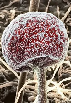
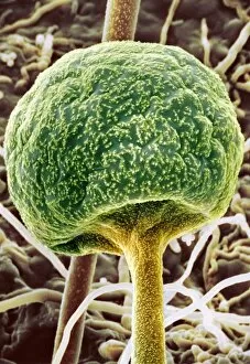
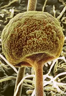
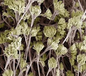
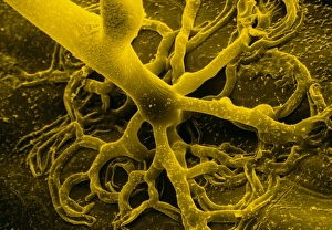
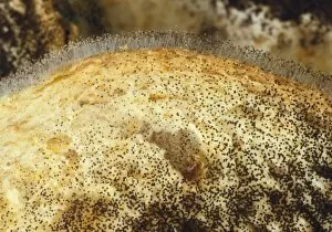
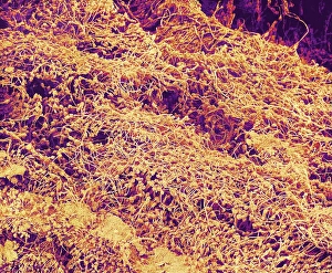
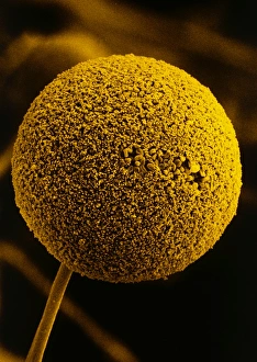
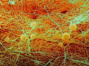
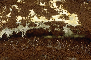
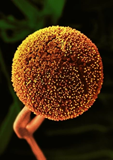
"Bread Mould: Unveiling the Intricate World of Fungal Growth" Delving into the microscopic realm, SEM (Scanning Electron Microscopy) images reveal fascinating details about bread mould. In ancient times, wooden moulds like the Mold for a Eulogia Bread were used to shape and bless loaves from the 600s to 900s. Captured under SEM C017 / 7127, SEM C017 / 7129, and SEM C017 / 7128, these intricate structures showcase the diverse forms of bread mould. The Light Micrograph (LM) at a staggering magnification of x850 exposes Rhizopus sporangia in all their glory – an awe-inspiring sight that spans across A4 dimensions. Penicillium fungus takes center stage in this captivating display with its unique characteristics highlighted through detailed SEM imagery. Its distinctive spores are showcased multiple times as they take on different shapes and sizes. As we explore further into this hidden world, more bread mould varieties come to light through additional SEM captures. Each image provides a glimpse into their complex structures and textures. However, it is not just their physical appearance that captivates us; it is also their role in nature's cycle. These resilient organisms play a crucial part in decomposition processes while contributing to nutrient recycling. Intriguingly, false-colored SEM images expose rhizoids - root-like structures that anchor bread mould firmly onto surfaces. This visual representation offers insight into how these fungi establish themselves within their environment. The study goes beyond mere curiosity; it holds significance in various fields such as food science and medicine. Understanding its growth patterns can aid in preventing spoilage or even unlocking potential medicinal properties found within certain strains. So next time you spot those fuzzy patches on your loaf or encounter mysterious growths elsewhere – remember that beneath our eyes lies an entire universe teeming with lifeforms like bread mould, silently shaping the world around us.