Fraud Blocker Skip to main content

White bread mould, SEM

White bread mould, SEM
In Stock
We accept all major payment options so you can pay for the things you need in the way that is most convenient for you
Framed Photos
Buy from AU$119.99
Photo Prints
Buy from AU$12.99
Jigsaw Puzzles
Buy from AU$89.99
Poster Prints
Buy from AU$24.99
Canvas Prints
Buy from AU$124.99
Fine Art Prints
Buy from AU$92.53
Metal Prints
Buy from AU$43.99
Cards
Buy from AU$12.99
Home Decor
Buy from AU$24.99

Science Photo Library Photo Prints and Wall Art

White bread mould, SEM

White bread mould. Coloured scanning electron micrograph (SEM) of the fruiting bodies of two types of mould growing on white bread. The moulds are Penicillium sp. and Mucor mucedo. Spores from these moulds circulate freely in the air. On a favourable medium, they germinate and form an extensive network of threads (hyphae) that absorb food for growth and spore production. The Mucor spores are borne in the sac-like bodies seen, which are called sporangia. The Penicillium spores, known as conidia, are borne directly on special hyphae called conidiophores. Magnification: x40 at 6x7cm size. Science Photo Library features Science and Medical images including photos and illustrations. © SUSUMU NISHINAGA/SCIENCE PHOTO LIBRARY

Media ID 6292343

Body Bread Mould Conidia Conidiophore Conidium Decay Decaying Eumycota Filament Filaments Fruiting Bodies Fungal Fungi Fungus Hypha Hyphae Mold Mould Mouldy Mycology Net Work Sporangia Sporangium

SECURE PAYMENTS
We accept all major payment options so you can pay for the things you need in the way that is most convenient for you

Made in Australia
Safe Shipping, Easy Returns

FREE PERSONALISATION
We are proud to offer a range of customisation including Cropping, Captions, and Filters. Zoom Cropping can be adjusted in the basket


EDITORS COMMENTS

This print showcases the intricate world of white bread mould, revealing a mesmerizing display of nature's decay and fungal growth. In this coloured scanning electron micrograph (SEM), we are presented with the fruiting bodies of two distinct types of mould thriving on a slice of white bread. The first type is Penicillium sp. , known for its characteristic blue-green appearance in other contexts. Its spores, called conidia, are directly borne on specialized hyphae called conidiophores. The second type is Mucor mucedo, whose spores are housed within sac-like structures known as sporangia. Both these moulds have their own unique way of spreading through the air; their microscopic spores circulate freely, waiting for a favorable environment to germinate and initiate an extensive network of threads called hyphae. These hyphae serve as conduits for absorbing nutrients necessary for growth and eventual spore production. In this image, we witness the remarkable complexity and beauty that lies within seemingly mundane objects like bread. The filaments intertwine to form an intricate net-like structure that serves as a testament to the resilience and adaptability of fungi in our ecosystem. With a magnification factor of 40 times at 6x7cm size, this SEM snapshot allows us to appreciate the hidden world teeming with life even in unexpected places such as decaying food items. It reminds us that there is always more than meets the eye when it comes to exploring nature's.


Framed Prints of White bread mould, SEM

Discover the fascinating world of microorganisms with Media Storehouse's Framed Prints. This captivating piece features a Coloured Scanning Electron Micrograph (SEM) image of White Bread Mould, showcasing the intricate structures of Penicillium sp. and Mucor mucedo as they grow. Each print is meticulously framed to preserve the image's vibrant colours and intricate details. Bring this unique and educational addition to your home or office, and start exploring the wonders of science.


Photo Prints of White bread mould, SEM

Discover the intriguing world of microorganisms with Media Storehouse's Photographic Prints. This captivating image, sourced from Science Photo Library, showcases the fascinating growth of Penicillium sp. and Mucor mucedo moulds on white bread. Viewed through the lens of a Scanning Electron Microscope (SEM), the vibrant colors and intricate details of these fruiting bodies bring a new perspective to the ordinary white bread. Add this unique print to your collection and ignite curiosity in the wonders of science and microbiology.


Poster Prints of White bread mould, SEM

Discover the intriguing world of microorganisms with our Media Storehouse Poster Prints. This captivating image showcases the beauty of White bread mould, as seen through the lens of a Scanning Electron Microscope from Science Photo Library. Witness the intricate details of Penicillium sp. and Mucor mucedo as they flourish on white bread in vibrant colors. Bring this mesmerizing microcosm into your home or office space, and ignite curiosity with every glance. Perfect for science enthusiasts, educators, and anyone with an appreciation for the unseen wonders of our world.


Jigsaw Puzzles of White bread mould, SEM

Discover the fascinating world of microorganisms with our Media Storehouse Jigsaw Puzzles! This intriguing puzzle features a captivating Coloured Scanning Electron Micrograph (SEM) image by Science Photo Library. Observe in detail as Penicillium sp. and Mucor mucedo moulds grow on white bread, revealing their unique textures and structures. A perfect addition to your collection for science enthusiasts, puzzle lovers, or anyone seeking a brain-teasing challenge with a difference. Assemble this enlightening puzzle and delve into the tiny yet wondrous world of microbes!

diamond
Made With High Grade Materials
thumb_up
Professional Quality Finish
package_2
Carefully Packed To Aid Safe Arrival

You May Also Like


Cactus spinesCactus spines
Add to Basket
Fairey Campania two-seat seaplaneFairey Campania two-seat seaplane
Add to Basket
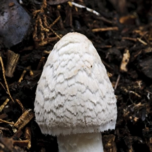
Magpie Fungus (Coprinopsis picacea) fruiting body, before veil has broken up to reveal dark cap underneathMagpie Fungus (Coprinopsis picacea) fruiting body, before veil has broken up to reveal dark cap underneath
Add to Basket
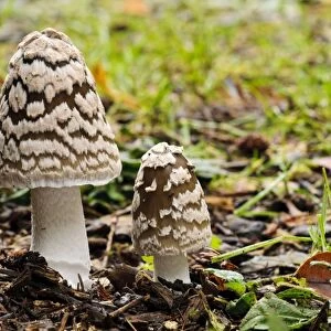
Magpie Fungus (Coprinopsis picacea) two fruiting bodies, Sir Harold Hillier Gardens, Romsey, Hampshire, EnglandMagpie Fungus (Coprinopsis picacea) two fruiting bodies, Sir Harold Hillier Gardens, Romsey, Hampshire, England
Add to Basket
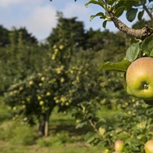
Cultivated Apple (Malus domestica) Howgate Wonder, close-up of fruit, on tree in orchard, England, AugustCultivated Apple (Malus domestica) Howgate Wonder, close-up of fruit, on tree in orchard, England, August
Add to Basket
Cultivated Apple (Malus domestica) close-up of developing fruit, Sweden, julyCultivated Apple (Malus domestica) close-up of developing fruit, Sweden, july
Add to Basket
Listening to The Old Curiosity Shop in the West. Illustration for Bret Hart sListening to The Old Curiosity Shop in the West. Illustration for Bret Hart s
Add to Basket
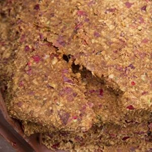
Cultivated Apple (Malus domestica) cake, left over from cider apple pressing at Benedictine monasteryCultivated Apple (Malus domestica) cake, left over from cider apple pressing at Benedictine monastery
Add to Basket
Cultivated Apple (Malus domestica) harvested fruit, cider apples ready for pressing at Benedictine monasteryCultivated Apple (Malus domestica) harvested fruit, cider apples ready for pressing at Benedictine monastery
Add to Basket
Picture No. 10884409Picture No. 10884409
Add to Basket
Picture No. 11050512Picture No. 11050512
Add to Basket
Achill Island, Co Galway, Ireland - The Dugort ColonyAchill Island, Co Galway, Ireland - The Dugort Colony
Add to Basket

Gallery Wall Inspiration

Yellow Slime Mold - growing on tree stump, Hessen, Germany
Riot at Mold in Flintshire
Bread mould, SEM
Detail of Christ's face from Michelangelo's Piet, artist's original cast. This work is on exhibit at the Academy of Art in Florence
Simurgh (Simorgh or Senmurw) - Azulejos - Faience de Kashan (Persia), period Ilkhanide
The death mask of Beethoven, molded by Josef Danhauser around twelve hours after Beethovens death. Ludwig van Beethoven, 1770 - 1827. German composer and pianist. From Ludwig van Beethoven, 1770 - 1827, Sein Leben in Bildern (His Life in Pictures)
Aspergillus
Molding Lead Soldiers
Scanning Electron Micrograph (SEM): Mycorrhiza fungus in plant root; Magnification x 700 (if printed A4, 29. 7 cm wide)
Former Liverpool Councillor Derek Hatton with his wife Shirley at Mold Crown Court for a
Slime mould (Lamproderma scintillans) super close up of 1mm tall sporangia
Slime mould (Physarum leucophaeum), two sporangia, close-up. Buckinghamshire, England, UK
Slime mould (Physarum album), dew droplets on two sporangia, close-up
Molds for the traditional glass-blowing workshop at Skansen. Stockholm, Sweden
Large group of Slime mould (Stemonitis flavogenita) sporangia changing to orange, beginning to mature on Oak (Quercus sp) log. Hertfordshire, England, UK. September. Focus Stacked
Pa. German Butter Mold, c. 1940. Creator: Albert J. Levone
Figure of Avalokiteshvara (gold)
Stag Plaque, 400-300 BC (gold, cast in shell mold)
Lloyd George laughing heartily over a joke with the Chaired Bard (next him on right), Mr
Women dressed in traditional clothing at the Eisteddfod at Mold, Flintshire
Lead Mine, Rhydymwyn, near Mold, Flintshire, Wales, 5th March 1938
Slime mould (Craterium minutum) tiny sporangia in various stages of development on tiny
Slime mould (Lamproderma scintillans) close up of sporangia growing on a Holly leaf
Slime mould (Physarum sp), dew droplets on sporangium, close-up
Slime mould (Cribraria argillacea), mature sporangia with cages that contain the spores
Slime mould (Comatricha nigra) sporangia with a range of colours
Sprinkler, Iran, 19th century. Creator: Unknown


© Copyright Media Storehouse, All Rights Reserved 2003 - 2024
Facebook     Pinterest     100% Payment Secure     Reviews IO