
Conidia Collection
Conidia, the microscopic spores produced by fungi, are a fascinating subject of study in the field of microbiology


All Professionally Made to Order for Quick Shipping
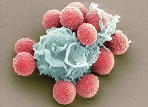
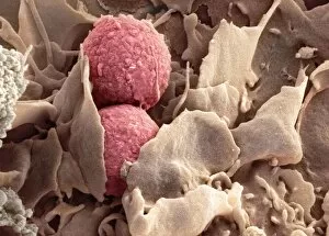
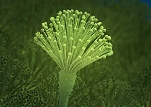
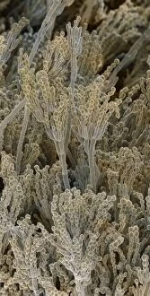
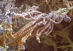
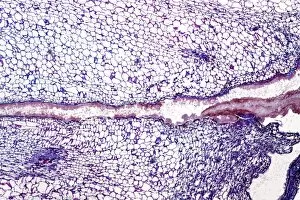
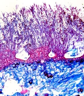
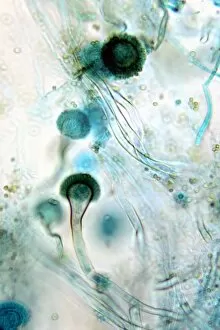
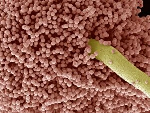
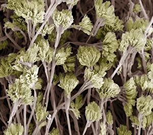
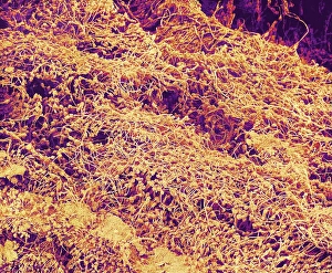
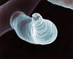
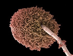
Conidia, the microscopic spores produced by fungi, are a fascinating subject of study in the field of microbiology. These tiny structures play a crucial role in the life cycle and dispersal of various fungal species. One captivating aspect is their interaction with other organisms. Through phagocytosis, immune cells engulf and destroy these fungal spores to protect the body from potential infections. Scanning Electron Micrographs (SEM) capture this intricate process, revealing the delicate dance between host and pathogen. In Picture No. 11675587, we witness phagocytosis of fungus spores under SEM. The image showcases the remarkable detail of this cellular event as immune cells engulf and eliminate invading fungal particles. Microscopic views also provide insight into specific fungal species like Stachybotrys chartarum. This black mold's they are be observed through a microscope, allowing scientists to better understand its structure and behavior. Another intriguing example is Entomophthora muscae parasitizing a fly. This parasitic fungus infects insects by releasing conidia onto their bodies, which then germinate and penetrate their hosts' tissues for sustenance. Artwork C013/4613 depicts Aspergillus fungus—a common genus known for its diverse conidial forms—highlighting both its beauty and scientific significance. Penicillium fungi are renowned for their antibiotic properties but are equally mesmerizing when viewed up close under an SEM lens. The detailed images reveal intricately shaped penicillium spores that aid in identification and classification within this group. Furthermore, SEM allows us to explore fruiting bodies or conidiophores—the reproductive structures responsible for producing conidia—in greater detail. Magnified at x7, 000 times actual size on A4 paper dimensions (29. 7 cm width), these images showcase Aspergillus sp. 's complex network of fruiting bodies with astonishing clarity.