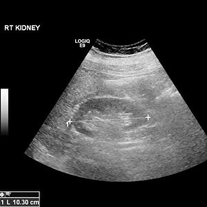
Penicillium mould, light micrograph

![]()
Wall Art and Photo Gifts from Science Photo Library
Penicillium mould, light micrograph
Penicillium mould. Light micrograph of a section through an orange rind infected with Penicillium sp. blue mould fungus, showing fungal hyphae and conidiophores (pink and purple) with spores (red). This saprophytic mould produces spores that germinate and pierce the rind of the orange. They then produce hyphae (tubular, purple), which infect the cells of the orange and suck out nutrients. The hyphae then pierce the rind with stalked conidiophores that produce rows of conidiospores on the outer surface of the rind, which are dispersed by the wind. Penicillium is used to produce penicillin, an antibiotic. Magnification: x70 when printed 10 centimetres wide
Science Photo Library features Science and Medical images including photos and illustrations
Media ID 6338791
© DR KEITH WHEELER/SCIENCE PHOTO LIBRARY
Citrus Sinensis Conidia Conidiophore Conidiophores Diseased Fruit Fungal Fungi Fungus Histological Histology Hypha Hyphae Infected Infection Micro Organism Micro Organisms Mold Orange Parasite Parasitic Parasitism Parasitology Penicillin Reproductive Structure Rind Saprophyte Saprophytic Spore Spores Structures Tissue Abnormal Cells Light Micrograph Light Microscope Micro Biology Microbiological Unhealthy
EDITORS COMMENTS
This print showcases the intricate world of Penicillium mould. The image reveals a section through an orange rind infected with the blue mould fungus, offering a glimpse into its parasitic nature. The pink and purple conidiophores stand out against the backdrop, adorned with vibrant red spores that are ready to be dispersed by the wind. The saprophytic mould's life cycle is unveiled in this snapshot. It begins with spores germinating and penetrating the orange rind, followed by the production of tubular hyphae that invade and extract nutrients from the fruit's cells. As if piercing through armor, stalked conidiophores emerge from these hyphae to release rows of conidiospores on the outer surface of the rind. Penicillium not only captivates us visually but also holds great significance in medicine as it is used to produce penicillin—an essential antibiotic. This light micrograph magnifies this microscopic wonder seventy times when printed at ten centimeters wide, allowing us to appreciate its beauty while delving into its biological intricacies. Through this stunning visual representation, we gain insight into how diseases can manifest even within seemingly healthy fruits like oranges. It serves as a reminder of nature's complexity and highlights our ongoing fascination with uncovering hidden wonders at a microscopic level.
MADE IN AUSTRALIA
Safe Shipping with 30 Day Money Back Guarantee
FREE PERSONALISATION*
We are proud to offer a range of customisation features including Personalised Captions, Color Filters and Picture Zoom Tools
SECURE PAYMENTS
We happily accept a wide range of payment options so you can pay for the things you need in the way that is most convenient for you
* Options may vary by product and licensing agreement. Zoomed Pictures can be adjusted in the Cart.