Fraud Blocker Skip to main content

Micro Biology Collection

Microbiology is a fascinating field that delves into the intricate world of microscopic organisms

Background imageMicro Biology Collection: Budding yeast cell

Budding yeast cell. Computer artwork of asection through a yeast ( Candida albicans )cell that is reproducing asexually. A daughtercell (top left) is budding from the parent cell

Background imageMicro Biology Collection: Avian flu virus

Avian flu virus, computer artwork. A virus is a tiny pathogenic particle comprising genetic material enclosed in a protein coat. The coat contains surface proteins (spikes)

Background imageMicro Biology Collection: Acrosphaera radiolarian, SEM

Acrosphaera radiolarian, SEM
Acrosphaera radiolarian. Coloured scanning electron micrograph (SEM) of the shell of a Acrosphaera sp. radiolarian. Radiolaria are single-celled protozoans that are found in marine plankton

Background imageMicro Biology Collection: Anthrax cultures, historical diagram

Anthrax cultures, historical diagram
Anthrax cultures. 1876 diagram of Bacillus anthracis (Anthrax) bacteria cultured by Robert Koch. Along with Louis Pasteur, Koch is considered the founder of modern medical bacteriology

Background imageMicro Biology Collection: Diatom, SEM

Diatom, SEM
Diatom. Coloured scanning electron micrograph (SEM) of the Arachnoidiscus sp. diatom. The diatoms are a group of photosynthetic, single- celled algae containing about 10, 000 species

Background imageMicro Biology Collection: Diatom, SEM

Diatom, SEM
Diatom. Coloured scanning electron micrograph (SEM) of a Stephanopyxis sp. diatom. Diatoms are single-celled photosynthetic algae, of which there are about 100, 000 species

Background imageMicro Biology Collection: E. coli bacteria, SEM

E. coli bacteria, SEM
E. coli bacteria. Coloured scanning electron micrograph (SEM) of Escherichia coli bacteria. These bacteria are a normal part of the intestinal flora in humans and other animals

Background imageMicro Biology Collection: Diatom, SEM

Diatom, SEM
Diatom. Coloured scanning electron micrograph (SEM) of a Triceratium morlandii diatom. Diatoms are single-celled photosynthetic algae, of which there are about 100, 000 species

Background imageMicro Biology Collection: PSCI2A-00015

PSCI2A-00015
Louis Pasteur in his laboratory. Hand-colored photogravure of an illustration by Albert Edelfelt

Background imageMicro Biology Collection: Herpes virus replicating

Herpes virus replicating, computer artwork.Viruses are only able to replicate in a host cell.The glycoprotein spikes in the virus envelope(green)

Background imageMicro Biology Collection: Embryonic stem cell and needle, SEM

Embryonic stem cell and needle, SEM
Embryonic stem cell and needle. Coloured scanning electron micrograph (SEM) of an embryonic stem cell (ESC) sitting in the eye of a needle

Background imageMicro Biology Collection: Calcareous phytoplankton, SEM

Calcareous phytoplankton, SEM
Calcareous phytoplankton. Coloured scanning electron micrograph (SEM) of the external mineralised structures (coccospheres) of small marine algal organisms called coccolithophores

Background imageMicro Biology Collection: Salmonella bacteria, SEM

Salmonella bacteria, SEM
Salmonella bacteria, coloured scanning electron micrograph (SEM). These Gram-negative rod-shaped bacteria can cause food poisoning when eaten in contaminated food

Background imageMicro Biology Collection: Coloured TEM of Yersinia pestis bacteria

Coloured TEM of Yersinia pestis bacteria
False colour transmission electron micrograph (SEM) of Yersinia pestis, a species of Gram negative, non- motile, capsulated

Background imageMicro Biology Collection: Candida fungus, SEM

Candida fungus, SEM
Candida fungus. Coloured scanning electron micrograph (SEM) of budding threads (hyphae) of a Candida fungus. The budding areas are where asexual reproduction is producing new fungus cells

Background imageMicro Biology Collection: HIV particle

HIV particle, computer artwork. HIV (human immunodeficiency virus) causes AIDS (acquired immune deficiency syndrome). The virus consists of an RNA genome

Background imageMicro Biology Collection: Avian flu virus

Avian flu virus, computer artwork. A virus is a tiny pathogenic particle comprising genetic material enclosed in a protein coat. The coat contains surface proteins (spikes)

Background imageMicro Biology Collection: Coloured SEM of mitochondria in ovarian cells

Coloured SEM of mitochondria in ovarian cells
Mitochondria. Coloured Scanning Electron Micro- graph (SEM) of mitochondria and smooth endoplasmic reticulum in an ovarian granulosa- lutein cell

Background imageMicro Biology Collection: Cell types, artwork

Cell types, artwork
Cell types. Cutaway artwork with a scale bar (upper right, in micrometres) showing the relative sizes of eukaryotic cells (those containing a nucleus) and prokaryotic cells (those lacking a nucleus)

Background imageMicro Biology Collection: Cell division, fluorescent micrograph

Cell division, fluorescent micrograph
Cell division. Immunofluorescent light micrograph of a human epithelial cell (centre) during the late anaphase stage of mitosis

Background imageMicro Biology Collection: Norovirus particles, TEM

Norovirus particles, TEM
Norovirus particles. Coloured transmission electron micrograph (TEM) of norovirus particles. Norovirus is a genus of RNA (ribonucleic acid)

Background imageMicro Biology Collection: Tuberculosis bacteria

Tuberculosis bacteria. Computer artwork of Mycobacterium tuberculosis bacteria. These Gram- positive rod-shaped bacteria cause the disease tuberculosis

Background imageMicro Biology Collection: Pseudomonas aeruginosa bacteria, SEM

Pseudomonas aeruginosa bacteria, SEM
Pseudomonas aeruginosa bacteria, coloured scanning electron micrograph (SEM). These Gram-negative rod-shaped bacteria are found in soil and water, and as normal flora in the human intestine

Background imageMicro Biology Collection: Spiral spore chain of Streptomyces bacteria

Spiral spore chain of Streptomyces bacteria

Background imageMicro Biology Collection: Dividing yeast cells, SEM

Dividing yeast cells, SEM
Dividing yeast cells. Coloured scanning electron micrograph (SEM) of Schizosaccharomyces pombe yeast cells dividing. S. pombe is a single-celled fungus that is studied widely as a model organism for

Background imageMicro Biology Collection: Dividing yeast cells, SEM

Dividing yeast cells, SEM
Dividing yeast cells. Coloured scanning electron micrograph (SEM) of Schizosaccharomyces pombe yeast cells dividing. S. pombe is a single-celled fungus that is studied widely as a model organism for

Background imageMicro Biology Collection: Coloured TEM of a Salmonella bacterium

Coloured TEM of a Salmonella bacterium
Salmonella sp. Coloured transmission electron micrograph (TEM) of a Salmonella sp. bacterium. The cell is rod-shaped with long hair-like flagellae

Background imageMicro Biology Collection: Bacteria infecting a macrophage, SEM

Bacteria infecting a macrophage, SEM
Bacteria infecting a macrophage. Coloured scanning electron micrograph (SEM) of Mycobacterium tuberculosis bacteria (purple) infecting a macrophage white blood cell

Background imageMicro Biology Collection: Flagellate bacteria

Flagellate bacteria. Computer artwork of a rod- shaped bacteria with polar flagella. Bacteria with a morphology similar to this include Helicobacter pylori and Salmonella sp

Background imageMicro Biology Collection: E. coli bacterium

E. coli bacterium
Escherichia coli. Coloured transmission electron micrograph (TEM) of an Escherichia coli bacterium. E. coli are Gram-negative bacilli (rod-shaped) bacteria

Background imageMicro Biology Collection: E. coli bacteria

E. coli bacteria
False-colour transmission electron micrograph of the bacterium Escherichia coli, a normal inhabitant of the human intestine

Background imageMicro Biology Collection: Staphylococcus aureus bacteria

Staphylococcus aureus bacteria
Staphylococcus aureus. Coloured Scanning Electron Micrograph (SEM) of Staphylococcus aureus bacteria. These Gram-positive bacteria cause skin infections

Background imageMicro Biology Collection: E. coli bacteria, SEM

E. coli bacteria, SEM
E. coli bacteria. Coloured scanning electron micrograph (SEM) of Escherichia coli bacteria (purple) taken from the small intestine of a child. E

Background imageMicro Biology Collection: Hepatitis B viruses

Hepatitis B viruses
Hepatitis B virus. Coloured transmission electron micrograph (TEM) of hepatitis B viruses (circles), a cause of liver inflammation

Background imageMicro Biology Collection: Salmonella bacteria, SEM

Salmonella bacteria, SEM
Salmonella bacteria, coloured scanning electron micrograph (SEM). Each rod-shaped structure is an individual bacterium. These bacteria occur mainly in human and animal intestines

Background imageMicro Biology Collection: Pseudomonas aeruginosa bacteria, SEM

Pseudomonas aeruginosa bacteria, SEM
Pseudomonas aeruginosa bacteria, coloured scanning electron micrograph (SEM). These Gram-negative rod-shaped bacteria are found in soil and water, and as normal flora in the human intestine

Background imageMicro Biology Collection: E. coli bacterium, TEM

E. coli bacterium, TEM
E. coli bacterium, coloured transmission electron micrograph (TEM). E. coli are Gram-negative rod-shaped bacteria that are part of the normal flora of the human gut

Background imageMicro Biology Collection: MRSA resistant Staphylococcus bacteria

MRSA resistant Staphylococcus bacteria
MRSA: resistant Staphylococcus bacteria. Coloured transmission electron micrograph (TEM) of a deadly cluster of MRSA Staphylococcus aureus bacteria

Background imageMicro Biology Collection: Sleeping sickness parasite

Sleeping sickness parasite
Sleeping sickness. Artwork of a trypanosome (Trypanosoma brucei) moving past human red blood cells in the blood. This protozoan is the cause of sleeping sickness (African trypanosomiasis)

Background imageMicro Biology Collection: Louis Pasteur, French microbiologist

Louis Pasteur, French microbiologist
Louis Pasteur (1822-1895), French microbiologist and chemist. Louis Pasteur proved the germ theory of disease, which holds that germs attack the body from the outside

Background imageMicro Biology Collection: Macrophage engulfing TB bacteria, SEM

Macrophage engulfing TB bacteria, SEM
Macrophage engulfing TB bacteria. Coloured scanning electron micrograph (SEM) of a macrophage white blood cell (purple) engulfing a tuberculosis (Mycobacterium tuberculosis) bacterium (pink)

Background imageMicro Biology Collection: Coronavirus particles, TEM

Coronavirus particles, TEM
Coronavirus particles. Coloured transmission electron micrograph (TEM) of three coronavirus particles. Different strains of coronavirus are responsible for diseases such as the common cold

Background imageMicro Biology Collection: Pasteurs Jubilee celebrations, 1892

Pasteurs Jubilee celebrations, 1892
Pasteurs Jubilee celebrations. Artwork of British surgeon Joseph Lister (in gown, walking up steps, 1827-1912) and French chemist Louis Pasteur (being approached by Lister)

Background imageMicro Biology Collection: Seed shrimp, light micrograph

Seed shrimp, light micrograph
Seed shrimp. Light micrograph of a seed shrimp (Cyclocypris sp.). The body of this freshwater crustacean is enclosed in two shells, which it can open and close using a special muscle

Background imageMicro Biology Collection: Phagocytosis of fungal spores, SEM

Phagocytosis of fungal spores, SEM
Phagocytosis of a fungus spore. Coloured scanning electron micrograph (SEM) of a neutrophil (blue) phagocytosing (engulfing and destroying) spores from the fungus Aspergillus fumigatus (red)

Background imageMicro Biology Collection: Hepatitis B viruses

Hepatitis B viruses

Background imageMicro Biology Collection: Neutrophil cell trapping bacteria, SEM

Neutrophil cell trapping bacteria, SEM
Neutrophil cell trapping bacteria. Coloured scanning electron micrograph (SEM) of bacteria (rod-shaped) being trapped by a neutrophil cell

Background imageMicro Biology Collection: Influenza virus, computer artwork

Influenza virus, computer artwork
Influenza virus. Computer artwork of an influenza (flu) virus. The virus consists of a core of RNA (ribonucleic acid) genetic material surrounded by a protein coat


All Professionally Made to Order for Quick Shipping

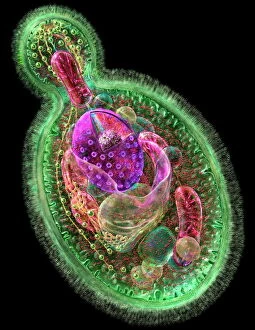
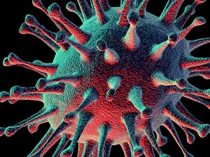
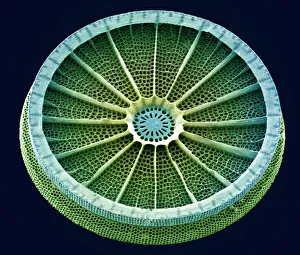
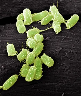
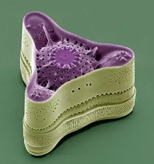
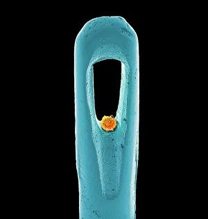
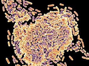
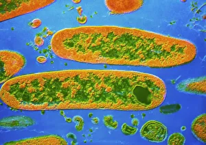
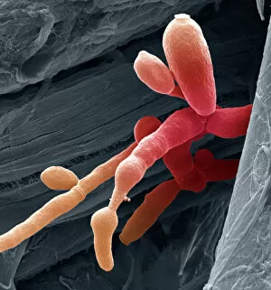
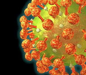
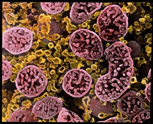
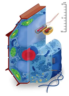
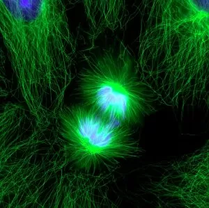
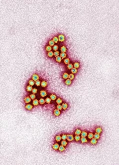
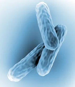
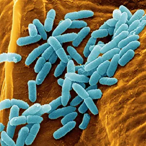
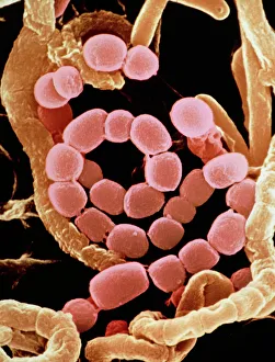
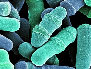
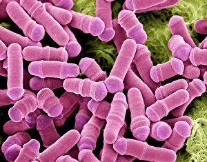
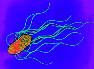
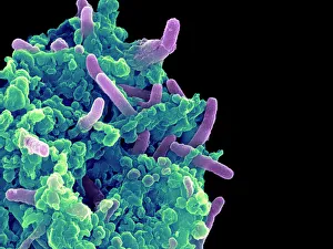
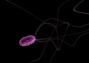
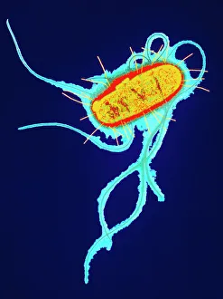
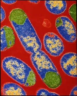
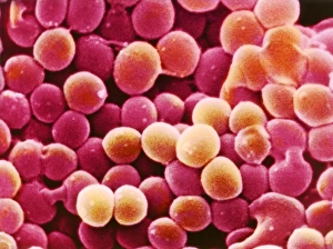
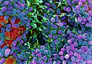
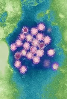
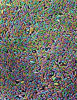
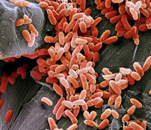
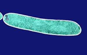
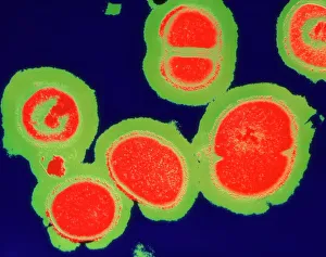
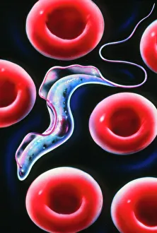
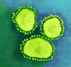
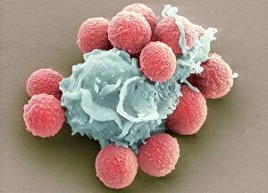
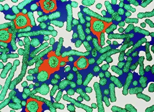
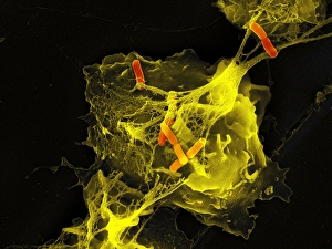
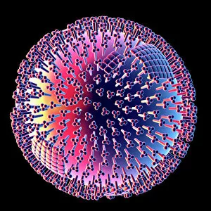
Microbiology is a fascinating field that delves into the intricate world of microscopic organisms. From budding yeast cells to calcareous phytoplankton, these tiny beings hold immense importance in our ecosystem. Take a closer look through the lens of a scanning electron microscope (SEM), and you'll be amazed by their intricate structures. One such example is the avian flu virus, which can cause devastating outbreaks among bird populations. Its unique shape and composition are revealed under SEM, highlighting its potential threat to both animals and humans alike. Diatoms, another group of microorganisms, showcase stunning beauty when observed through SEM. Their delicate silica shells form intricate patterns that resemble works of art. These diatoms play a crucial role in marine ecosystems as primary producers. E. coli bacteria, often associated with foodborne illnesses, reveal their rod-shaped structure under SEM. Understanding their morphology helps scientists develop strategies to combat infections caused by this notorious bacterium. The PSCI2A-00015 embryonic stem cell captured under SEM demonstrates its incredible regenerative capabilities. This versatile cell type holds great promise for future medical advancements and treatments. Salmonella bacteria also come into focus under SEM, displaying their flagella-covered surface responsible for motility and infection transmission. Studying these pathogens aids in developing effective prevention measures against salmonellosis. Intriguingly colored TEM images unveil Yersinia pestis bacteria responsible for causing deadly diseases like bubonic plague throughout history. Unveiling their detailed structure allows researchers to better understand how they function within host organisms. Another captivating diatom species reveals itself through SEM imaging - showcasing nature's artistic side once again. These single-celled algae contribute significantly to global carbon fixation while providing habitats for countless aquatic creatures. Candida fungus captures attention with its filamentous appearance when viewed at high magnification using SEM techniques. This opportunistic pathogen can cause various infections in humans ranging from mild oral thrush to life-threatening systemic infections.