Home > Science > SEM
Neutrophil cell trapping bacteria, SEM

![]()
Wall Art and Photo Gifts from Science Photo Library
Neutrophil cell trapping bacteria, SEM
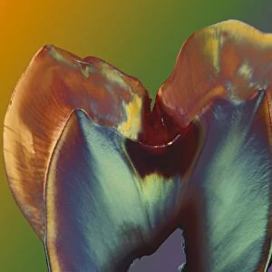
Neutrophil cell trapping bacteria. Coloured scanning electron micrograph (SEM) of bacteria (rod-shaped) being trapped by a neutrophil cell. The neutrophil cell (a type of white blood cell) has trapped the bacteria with extruded material that forms a net-like structure called a NET (neutrophil extracellular trap). This method of cellular defence was first discovered in 2004. These are Shigella sp. bacteria, one of the causes of dysentery, a severe intestinal inflammation
Science Photo Library features Science and Medical images including photos and illustrations
Media ID 6421262
© SCIENCE PHOTO LIBRARY
Antigen Bacilli Bacillus Bacteria Bacterial Bacteriology Bacterium Cytology Defence Defending Digestion Digestive Dysentery False Colour Immune Immune System Immunology Intestinal Neutrophil Pathological Pathology Shigella System Trapping White Blood Cell Cells Eliminating False Coloured Micro Biology Microbiological
FEATURES IN THESE COLLECTIONS
EDITORS COMMENTS
This print from Science Photo Library showcases the intricate and fascinating world of cellular defense within our immune system. In this colored scanning electron micrograph (SEM), we witness a remarkable scene where a neutrophil cell, a type of white blood cell, traps bacteria using an extruded material that forms a net-like structure called a neutrophil extracellular trap (NET). The image reveals rod-shaped Shigella sp. bacteria being ensnared by the vigilant neutrophil cell. These particular bacteria are known to cause dysentery, a severe inflammation of the intestines. The NET created by the neutrophil acts as an effective method for trapping and eliminating these harmful pathogens. First discovered in 2004, this unique mechanism of cellular defense highlights the complexity and sophistication of our immune system's response to bacterial invasion. It provides us with valuable insights into how our bodies combat infections and protect against diseases. Through false coloring techniques applied during scanning electron microscopy, we can appreciate the intricacies of this microscopic battle taking place within our own bodies. This stunning image not only serves as an invaluable resource for medical professionals studying bacteriology, cytology, pathology, immunology, and microbiology but also offers viewers an awe-inspiring glimpse into the hidden wonders that lie within us. As we marvel at this mesmerizing photograph captured by Science Photo Library's skilled photographers and scientists alike, let it serve as a reminder of both the fragility and resilience inherent in our human anatomy.
MADE IN AUSTRALIA
Safe Shipping with 30 Day Money Back Guarantee
FREE PERSONALISATION*
We are proud to offer a range of customisation features including Personalised Captions, Color Filters and Picture Zoom Tools
SECURE PAYMENTS
We happily accept a wide range of payment options so you can pay for the things you need in the way that is most convenient for you
* Options may vary by product and licensing agreement. Zoomed Pictures can be adjusted in the Cart.