
Pathological Collection
"Unveiling the Pathological Mysteries: Exploring the Intricacies of Disease" Delving into the realm of pathology, where secrets lie beneath the surface


All Professionally Made to Order for Quick Shipping
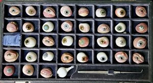
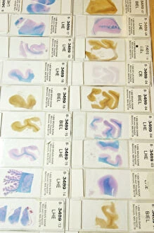
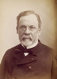
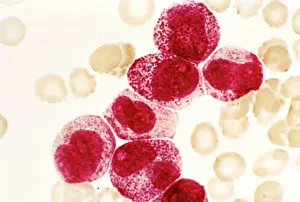
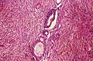
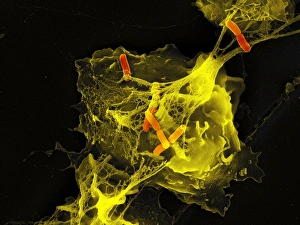
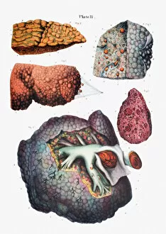
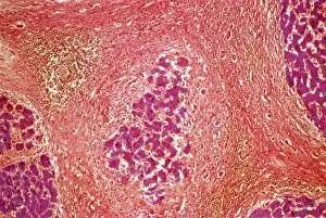
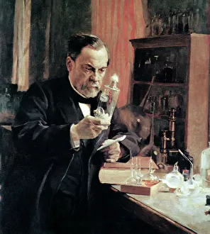
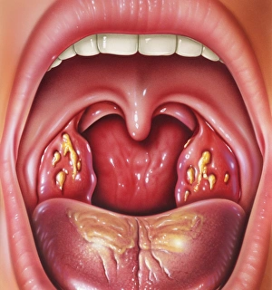
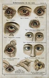
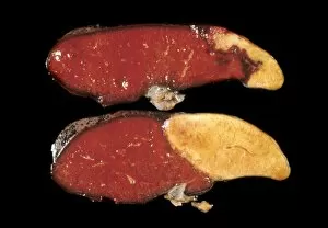
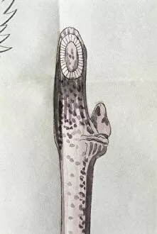
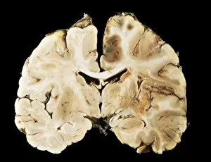
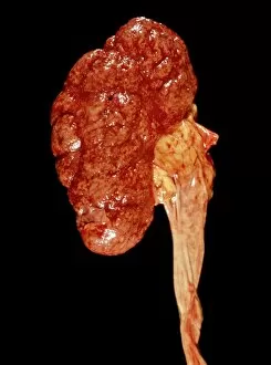
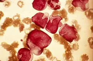
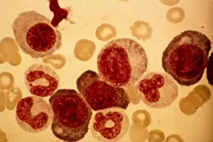
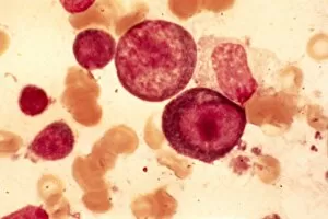
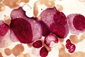
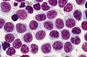
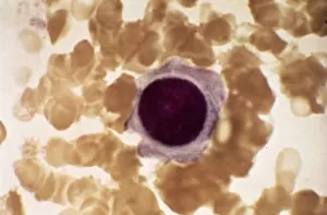
"Unveiling the Pathological Mysteries: Exploring the Intricacies of Disease" Delving into the realm of pathology, where secrets lie beneath the surface, we encounter a fascinating array of specimens. From a set of glass eyeballs to human brain microscope slides, each holds a story waiting to be told. In this captivating journey, we pay homage to Louis Pasteur, the pioneering French microbiologist whose discoveries revolutionized medicine. His relentless pursuit for knowledge paved the way for understanding diseases at their core. Peering through microscopes reveals Dohle bodies in blood cells and acute promyelocytic leukemia micrographs. These hauntingly beautiful images remind us of the intricate battles waged within our bodies against formidable foes. Ovarian cancer comes into focus with its light micrograph C015/7103 showcasing its devastating impact on delicate tissues. As we witness liver cirrhosis unfold before our eyes through another light micrograph, we are reminded of how vital it is to protect this resilient organ from harm. Cystic fibrosis takes center stage as we explore its effects on lung tissue and delve deeper into understanding this complex genetic disorder that affects countless lives worldwide. Returning once again to Louis Pasteur's legacy, his contributions resonate as osteoporotic bone captures our attention. We reflect on his tireless efforts in unraveling mysteries that continue to shape modern medicine today. Finally, herpes simplex viruses reveal themselves under an electron microscope - tiny yet potent agents causing widespread discomfort and reminding us of humanity's constant battle against infectious diseases. Through these glimpses into pathology's intricate world, we gain insight into both the beauty and fragility inherent in life itself. Each image serves as a testament to human resilience and scientific progress - guiding us towards better prevention strategies and treatments for those affected by these pathological conditions.