Fraud Blocker Skip to main content

Pathological Collection (page 4)

"Unveiling the Pathological Mysteries: Exploring the Intricacies of Disease" Delving into the realm of pathology, where secrets lie beneath the surface

Background imagePathological Collection: Human brain dissected in half

Human brain dissected in half. A human brain dissected into right and left hemispheres. It has been donated for medical research at the Harvard Brain and Tissue Resource Centre, USA

Background imagePathological Collection: Bacterial infection of nail, SEM

Bacterial infection of nail, SEM
Bacterial infection of a nail. Coloured scanning electron micrograph (SEM) of a nail with a bacterial infection (paronychia)

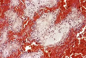
Background imagePathological Collection: Bleeding in emphysema

Bleeding in emphysema
Internal bleeding in lung tissue due to pulmonary emphysema, light micrograph. The bleeding is seen as numerous red blood cells (red)

Background imagePathological Collection: Studying the brain, conceptual image

Studying the brain, conceptual image

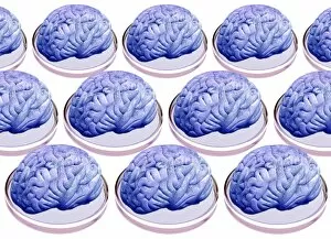
Background imagePathological Collection: Brains in petri dishes, conceptual image

Brains in petri dishes, conceptual image
Brains in petri dishes, conceptual artwork

Background imagePathological Collection: Neutrophil cell and bacteria, SEM

Neutrophil cell and bacteria, SEM
Neutrophil cell and bacteria. Coloured scanning electron micrograph (SEM) of bacteria (rod-shaped) and part of a neutrophil cell (lower left)

Background imagePathological Collection: Macrophage cell engulfing bacteria, TEM

Macrophage cell engulfing bacteria, TEM
Macrophage cell engulfing bacteria. Coloured transmission electron micrograph (TEM) of bacteria (centre, rod-shaped) inside a macrophage cell

Background imagePathological Collection: Back deformities

Back deformities, historical anatomical artwork. These patients all have spinal deformities leading to abnormal curvatures of the back

Background imagePathological Collection: Aortic aneurysm

Aortic aneurysm. Artwork from Jean Cruveilhiers medical textbook Anatomie pathalogique du corps humain, published 1835. The two large illustrations are views of an aortic aneurysm (brown, centre)

Background imagePathological Collection: AIDS in the UK

AIDS in the UK, conceptual image. Computer artwork of red ribbons overlain on a map of the UK. These ribbons are worn to show support for the cause of AIDS prevention

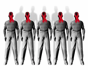
Background imagePathological Collection: People with AIDS

People with AIDS, conceptual image. Computer artwork of red ribbons overlaid on the faces of people with AIDS. These ribbons are worn to show support for the cause of AIDS prevention

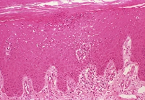
Background imagePathological Collection: Psoriasis, light micrograph

Psoriasis, light micrograph
Psoriasis. Light micrograph of a section through a psoriasis pustule. Psoriasis is a chronic skin disease causing inflammation and scaly lesions

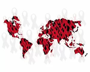
Background imagePathological Collection: World AIDS epidemic

World AIDS epidemic, conceptual image. Computer artwork of red ribbons overlain on a map of the world. These ribbons are worn to show support for the cause of AIDS prevention

Background imagePathological Collection: Edme Vulpian, French neurologist

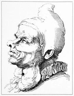
Edme Vulpian, French neurologist
Edme Felix Alfred Vulpian (1826-1887), French neurologist. Vulpian was born and educated in Paris. His doctoral thesis (1853) was on the origin of cranial nerves III to X

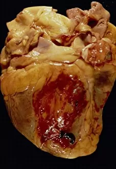
Background imagePathological Collection: Ruptured heart

Ruptured heart. Gross specimen of a heart that has ruptured in the parietal region (dark blood clots, lower centre, and red region, centre)

Background imagePathological Collection: Obsessive compulsive disorder

Obsessive compulsive disorder. Computer artwork of a DNA (deoxyribonucleic acid) autoradiogram spelling out OCD (obsessive compulsive disorder)

Background imagePathological Collection: Typhoid nodules, light micrograph

Typhoid nodules, light micrograph
Typhoid nodules, coloured light micrograph. Section through a lymph node of a patient with typhoid, showing macrophages (large cells, pink) forming typhoid nodules

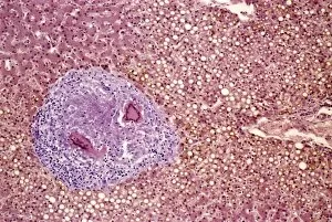
Background imagePathological Collection: Liver tuberculosis, light micrograph

Liver tuberculosis, light micrograph
Liver tuberculosis. Coloured light micrograph of a section through the liver of a patient with miliary tuberculosis (TB). A tubercle, a nodular lesion of infected dead tissue, is seen at left

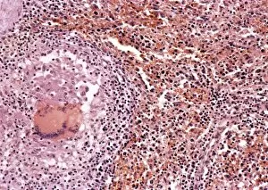
Background imagePathological Collection: Miliary tuberculosis, light micrograph

Miliary tuberculosis, light micrograph
Miliary tuberculosis. Light micrograph of a section through a single miliary tubercle (left) in a patients lung. Tubercles are nodular lesions of infected dead tissue that arise from tuberculosis (TB)

Background imagePathological Collection: Demyelinated nerve in multiple sclerosis

Demyelinated nerve in multiple sclerosis. Coloured transmission electron micrograph (TEM) of a section through an axon (a structure that transmits nerve impulses) that has lost its myelin sheath

Background imagePathological Collection: Multiple sclerosis, SEM

Multiple sclerosis, SEM
Multiple sclerosis. Coloured scanning electron micrograph (SEM) of microglial cells (round) ingesting oligodendrocytes (branched). This is the process thought to occur in multiple sclerosis (MS)

Background imagePathological Collection: Systemic lupus erythematosus

Systemic lupus erythematosus. Light micrograph of a section through a glomerulus (blood filtration structure) from kidney tissue in a case of SLE

Background imagePathological Collection: Coronary thrombosis, light micrograph

Coronary thrombosis, light micrograph
Coronary thrombosis. Light micrograph of a section through a thrombus (blood clot) in the coronary artery of the heart. The clot is packed full of red blood cells (red)

Background imagePathological Collection: Giovanni Battista Morgagni, anatomist

Giovanni Battista Morgagni, anatomist

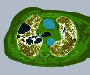
Background imagePathological Collection: Lung fibrosis, CT scan

Lung fibrosis, CT scan
Lung fibrosis. Image 2 of 3. Coloured computed tomography (CT) scan through the chest of a patient with lung (pulmonary) fibrosis

Background imagePathological Collection: Creutzfeldt-Jakob diseased brain

Creutzfeldt-Jakob diseased brain
Creutzfeldt-Jakob disease (CJD). Computer artwork based on a light micrograph of a section through a human brain exhibiting CJD

Background imagePathological Collection: Crohns disease, X-ray

Crohns disease, X-ray

Background imagePathological Collection: Skin inflammation, light micrograph

Skin inflammation, light micrograph
Skin inflammation. Light micrograph of a section through skin showing inflamed capillaries (lower left and upper right) caused by capillaritis, also known as pigmented purpurea

Background imagePathological Collection: Bilharzia infection, ureter tissue

Bilharzia infection, ureter tissue
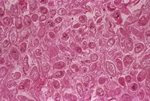
Bilharzia infection. Light micrograph of human ureter tissue that contains numerous eggs from Schistosoma flukes. The ureters are tubes that carry urine from the kidneys to the bladder

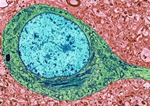
Background imagePathological Collection: Alzheimers disease brain cell, TEM

Alzheimers disease brain cell, TEM
Alzheimers disease brain cell. Coloured transmission electron micrograph (TEM) of a neurofibrillary tangle in a nerve cell from the brain of a patient with Alzheimers disease

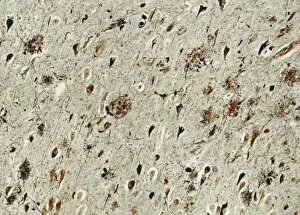
Background imagePathological Collection: Alzheimers disease brain tissue

Alzheimers disease brain tissue, light micrograph. Two characteristic features of Alzheimers disease are seen here; neurofibrillary tangles (dark teardrop shapes)

Background imagePathological Collection: Amyloidosis, TEM

Amyloidosis, TEM
Amyloidosis. Coloured transmission electron micrograph (TEM) of a section through tissue with abnormal deposoits of amyloid protein (green)

Background imagePathological Collection: Avian influenza virus, TEM

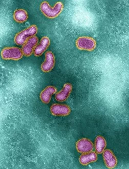
Avian influenza virus, TEM
Avian influenza virus, type A strain H5N1, coloured transmission electron micrograph (TEM). This virus was isolated in Vietnam, during the avian flu outbreak in early 2004

Background imagePathological Collection: Simian AIDS viruses, TEM

Simian AIDS viruses, TEM
Simian AIDS virus particles, coloured transmission electron micrograph (TEM). These virus particles (small, round) are bursting out of a cell (across bottom) after using the cell to replicate

Background imagePathological Collection: West Nile virus, TEM

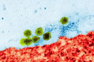
West Nile virus, TEM
West Nile virus particles. Coloured transmission electron micrograph (TEM) of West Nile virus (WNV) particles (green) next to a host cell (red)

Background imagePathological Collection: Vaccinia viruses, TEM

Vaccinia viruses, TEM
Vaccinia virus particles. Coloured transmission electron micrograph (TEM) of sectioned vaccinia virus particles. The genetic cores (red) are covered by membrane layers (green)

Background imagePathological Collection: Pig retrovirus, TEM

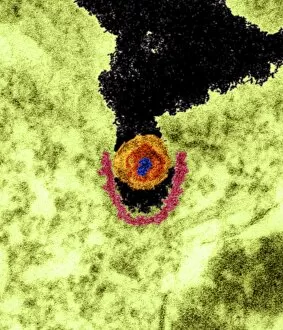
Pig retrovirus, TEM
Pig retrovirus. Coloured transmission electron micrograph (TEM) of porcine endogenous retrovirus (PERV) in infected tissue. The retrovirus is the orange/red/blue structure at centre

Background imagePathological Collection: Marie-Francois Bichat, pathologist

Marie-Francois Bichat, pathologist
Marie-Francois Xavier Bichat (1771-1802), French pathologist and founder of modern histology. Bichat noted that various organs consist of several components, or tissues

Background imagePathological Collection: E. coli bacteria colony, light micrograph

E. coli bacteria colony, light micrograph
Escherichia coli bacteria colonies growing on a nutrient substrate, differential interference contrast light micrograph. E

Background imagePathological Collection: Inflamed lymph gland, light micrograph

Inflamed lymph gland, light micrograph

Background imagePathological Collection: Scabies-affected skin, light micrograph

Scabies-affected skin, light micrograph

Background imagePathological Collection: Asthma pathology, artwork

Asthma pathology, artwork
Asthma pathology. Computer artwork showing cross-sections through a normal airway (right) and an asthmatic, constricted airway (left)

Background imagePathological Collection: Neurofibromatosis, 17th century artwork

Neurofibromatosis, 17th century artwork
Neurofibromatosis. 17th century artwork of a man with neck deformities (neurofibromas) due to neurofibromatosis. These tumours arise from the fibrous coverings of nerves

Background imagePathological Collection: Parasite and cells

Parasite and cells
Concept image. An aggresive unknown Parasite has entered a cell and consumes it from within

Background imagePathological Collection: Macrophage engulfing pathogen, artwork

Macrophage engulfing pathogen, artwork
Macrophage engulfing pathogen. Artwork of a macrophage cell (pink) engulfing a pathogen (green), a process known as phagocytosis. The pathogen will be destroyed once it has been engulfed

Background imagePathological Collection: Macrophage engulfing pathogens, artwork

Macrophage engulfing pathogens, artwork
Macrophage engulfing pathogens. Artwork of a macrophage cell (pink) engulfing pathogens (green), a process known as phagocytosis. The pathogens will be destroyed once they have been engulfed
Background imagePathological Collection: Virus particle, artwork

Virus particle, artwork. The spikes on this virus particle are surface proteins that help the particle attach to cell membranes

Background imagePathological Collection: Legionella bacteria, SEM

Legionella bacteria, SEM
Legionella bacteria. Coloured scanning electron micrograph (SEM) of Legionella pneumophila bacteria, the cause of Legionnaires disease


All Professionally Made to Order for Quick Shipping

"Unveiling the Pathological Mysteries: Exploring the Intricacies of Disease" Delving into the realm of pathology, where secrets lie beneath the surface, we encounter a fascinating array of specimens. From a set of glass eyeballs to human brain microscope slides, each holds a story waiting to be told. In this captivating journey, we pay homage to Louis Pasteur, the pioneering French microbiologist whose discoveries revolutionized medicine. His relentless pursuit for knowledge paved the way for understanding diseases at their core. Peering through microscopes reveals Dohle bodies in blood cells and acute promyelocytic leukemia micrographs. These hauntingly beautiful images remind us of the intricate battles waged within our bodies against formidable foes. Ovarian cancer comes into focus with its light micrograph C015/7103 showcasing its devastating impact on delicate tissues. As we witness liver cirrhosis unfold before our eyes through another light micrograph, we are reminded of how vital it is to protect this resilient organ from harm. Cystic fibrosis takes center stage as we explore its effects on lung tissue and delve deeper into understanding this complex genetic disorder that affects countless lives worldwide. Returning once again to Louis Pasteur's legacy, his contributions resonate as osteoporotic bone captures our attention. We reflect on his tireless efforts in unraveling mysteries that continue to shape modern medicine today. Finally, herpes simplex viruses reveal themselves under an electron microscope - tiny yet potent agents causing widespread discomfort and reminding us of humanity's constant battle against infectious diseases. Through these glimpses into pathology's intricate world, we gain insight into both the beauty and fragility inherent in life itself. Each image serves as a testament to human resilience and scientific progress - guiding us towards better prevention strategies and treatments for those affected by these pathological conditions.