
Bacillus Collection
"Bacillus: Unveiling the Microscopic World of Bacteria" Delving into the microscopic realm, we encounter a diverse array of bacilli


All Professionally Made to Order for Quick Shipping
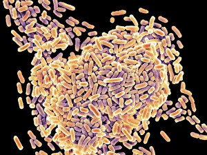
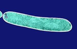
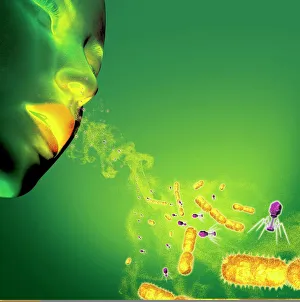
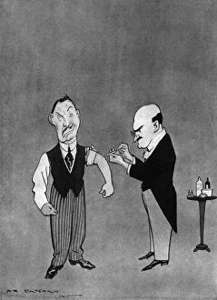
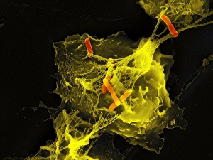
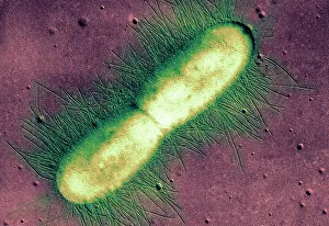
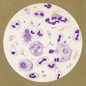
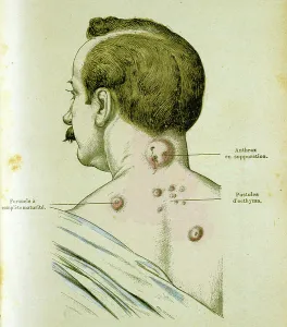
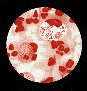
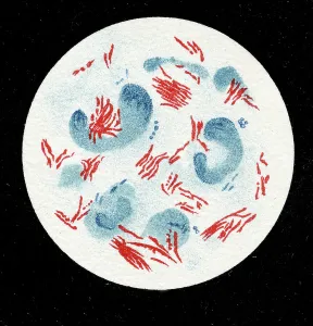
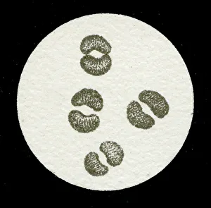
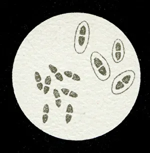
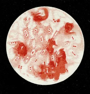
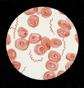
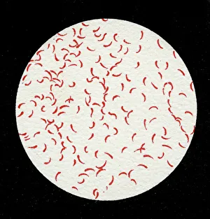
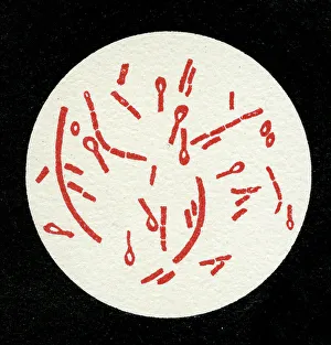
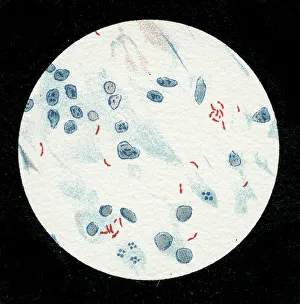
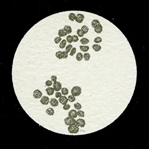
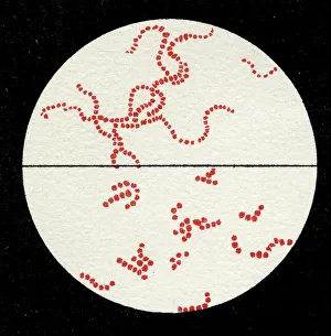
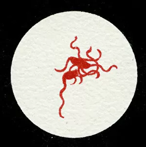
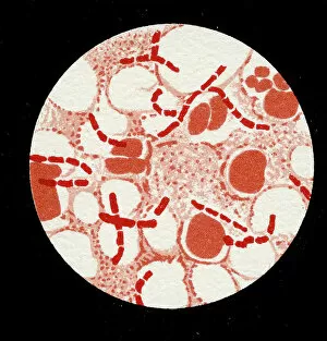
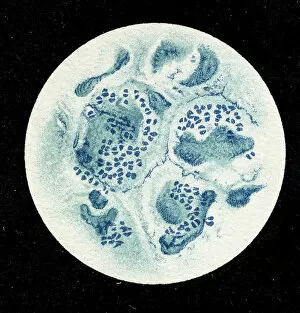
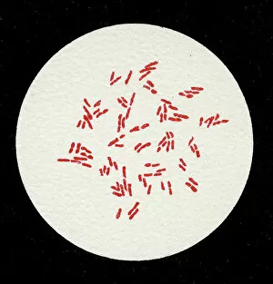
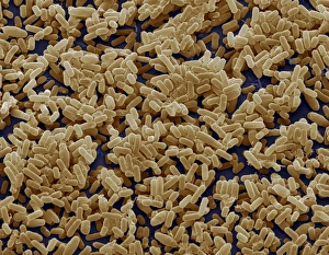
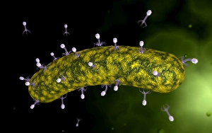
"Bacillus: Unveiling the Microscopic World of Bacteria" Delving into the microscopic realm, we encounter a diverse array of bacilli. From the notorious Salmonella bacteria, responsible for foodborne illnesses, to the E. Coli bacterium that can cause severe gastrointestinal infections – these tiny organisms wield significant impact on our health. Through scanning electron microscopy (SEM), we witness astonishing visuals of bacteria found on unexpected surfaces like mobile phones. It serves as a stark reminder to keep our devices clean and germ-free. In H. M. Bateman's intriguing artwork titled "Do you want some?", an artistic representation captures the essence of bacterial transmission through sneezing - highlighting how easily infections can spread in close quarters. Further exploring with transmission electron microscopy (TEM), we observe intricate details of an E. coli bacterium, revealing its complex structure and mechanisms at play. The tuberculosis bacillus emerges as another formidable foe within this microbial world. Artwork depicting both tuberculosis bacteria and bacteriophages showcases their interplay in this ongoing battle against infectious diseases. Taking us back in time, lithographs from 1906 showcase colonies of Haemophilus influenzae and Mycobacterium leprae - providing historical context to our understanding of these pathogens' existence long before modern scientific advancements. Lastly, SEM imagery unveils a captivating view of Salmonella typhimurium bacteria thriving amidst their surroundings; a testament to their resilience and adaptability even under extreme conditions. As we explore the fascinating world of bacilli, it becomes evident that these microorganisms hold immense power over human health. Understanding them better equips us in combating infectious diseases while appreciating their complexity within nature's intricate tapestry.