Fraud Blocker Skip to main content

Intestinal Collection

Intestinal health is a complex and fascinating topic that involves various organisms and structures

Background imageIntestinal Collection: Dog tapeworm head, SEM

Dog tapeworm head, SEM
Dog tapeworm head. Coloured scanning electron micrograph (SEM) of the head (scolex) of a dog tapeworm (Taenia pisiformis)

Background imageIntestinal Collection: Small intestine, SEM

Small intestine, SEM
Small intestine. Coloured scanning electron micrograph (SEM) of a biopsy from the small intestines of a child. The small intestine runs from the stomach to the large intestine

Background imageIntestinal Collection: E. coli bacteria, SEM

E. coli bacteria, SEM
E. coli bacteria. Coloured scanning electron micrograph (SEM) of Escherichia coli bacteria (purple) taken from the small intestine of a child. E

Background imageIntestinal Collection: Salmonella bacteria, SEM

Salmonella bacteria, SEM
Salmonella bacteria, coloured scanning electron micrograph (SEM). Each rod-shaped structure is an individual bacterium. These bacteria occur mainly in human and animal intestines

Background imageIntestinal Collection: E. coli bacterium, TEM

E. coli bacterium, TEM
E. coli bacterium, coloured transmission electron micrograph (TEM). E. coli are Gram-negative rod-shaped bacteria that are part of the normal flora of the human gut

Background imageIntestinal Collection: Deer anatomy, artwork

Deer anatomy, artwork
Deer anatomy. Computer artwork showing the internal anatomy of a deer (family Cervidae). Anatomical features shown here include the skeleton, lungs, intestines, bladder

Background imageIntestinal Collection: Human digestive system, artwork

Human digestive system, artwork. At top, food from the mouth forms a bolus that is swallowed down the oesophagus. At centre, the bolus is about to enter the stomach (arrow)

Background imageIntestinal Collection: Neutrophil cell trapping bacteria, SEM

Neutrophil cell trapping bacteria, SEM
Neutrophil cell trapping bacteria. Coloured scanning electron micrograph (SEM) of bacteria (rod-shaped) being trapped by a neutrophil cell

Background imageIntestinal Collection: Intestinal microvilli, TEM

Intestinal microvilli, TEM
Intestinal microvilli, coloured transmission electron micrograph (TEM). The microvilli are finger-like projections (seen here in circular cross-section)

Background imageIntestinal Collection: Cholera bacteria, artwork

Cholera bacteria, artwork
Cholera bacteria. Artwork of Vibrio cholerae bacteria in the small intestine. These Gram-negative rod-shaped bacteria have a single polar flagellum (tail-like structure)

Background imageIntestinal Collection: Cholera toxin, molecular model

Cholera toxin, molecular model
Cholera toxin. Molecular model of the secondary structure of cholera enterotoxin (intestinal toxin). The molecule consists of two subunits, A (top) and B (bottom)

Background imageIntestinal Collection: An advertisement for Tamar Indien Grillon a refreshing fruit lozenge to relieve constipation

An advertisement for Tamar Indien Grillon a refreshing fruit lozenge to relieve constipation and other gastric
5311141 An advertisement for Tamar Indien Grillon a refreshing fruit lozenge to relieve constipation and other gastric and intestinal troubles

Background imageIntestinal Collection: Cholera Morbus, from Relacao, Historica, Statistica e Medica da Cholera-Morbus

Cholera Morbus, from Relacao, Historica, Statistica e Medica da Cholera-Morbus
CHT236344 Cholera Morbus, from Relacao, Historica, Statistica e Medica da Cholera-Morbus by Francisco d Assis de Souza Vaz, 1833 (coloured engraving) by French School

Background imageIntestinal Collection: Parasitic tapeworm of the genus Taenia

Parasitic tapeworm of the genus Taenia. Copperplate engraving by Milton from Abraham Rees Cyclopedia or Universal Dictionary of Arts, Sciences and Literature, Longman, Hurst, Rees, Orme and Brown

Background imageIntestinal Collection: Intestinal worms

Intestinal worms: Pork tapeworm, Taenia solium 1, a, b, c, and human polycephalus, Polycephalus hominis 2, d, e. Handcoloured copperplate engraving from Friedrich Johann Bertuchs Bilderbuch fur

Background imageIntestinal Collection: The external surface of the intestines infected with cholera

The external surface of the intestines infected with cholera
CHT236338 The external surface of the intestines infected with cholera, from Anatomie Pathologique du Cholera Morbus, by Nikolai Ivanovich Pirogov (1810-81) published in St

Background imageIntestinal Collection: Simple intestinal massage (engraving)

Simple intestinal massage (engraving)
3097155 Simple intestinal massage (engraving) by German School, (19th century); Private Collection; (add.info.: Simple intestinal massage. Circular motions around the navel)

Background imageIntestinal Collection: Female skeleton and organs, artwork

Female skeleton and organs, artwork
Female skeleton and organs and blood vessels, artwork

Background imageIntestinal Collection: Giardia lamblia protozoa, micrograph

Giardia lamblia protozoa, micrograph
Giardia lamblia protozoa, light micrograph. These single-celled parasitic protozoa infect the intestinal tract and are most common in tropical regions

Background imageIntestinal Collection: Intestinal villi, SEM

Intestinal villi, SEM
Intestinal villi. Coloured scanning electron micrograph (SEM) of a section through a part of the small intestine, showing the villi that cover its inner surface

Background imageIntestinal Collection: Pancreatic acinar cell

Pancreatic acinar cell. Transmission electron micrograph (TEM) of a section through an enzyme-secreting acinar cell in the human pancreas, showing part of the nucleus (round, far left)

Background imageIntestinal Collection: Vagus nerve anatomy, artwork C018 / 0300

Vagus nerve anatomy, artwork C018 / 0300
Vagus nerve anatomy. Artwork of a human body from the side, showing the internal anatomy and the location of the vagus nerve (yellow)

Background imageIntestinal Collection: Early 19th Century enema syringe

Early 19th Century enema syringe. Pewter syringe used to administer enemas. Made in 1820

Background imageIntestinal Collection: Earthworm, transverse section

Earthworm, transverse section
Earthworm. Light micrograph of a transverse section through the body of a round segmented earthworm (Lumbricus terrestris) in the intestinal region

Background imageIntestinal Collection: Cow anatomy, artwork

Cow anatomy, artwork
Cow anatomy. Computer artwork showing the internal anatomy of a domesticated cow (Bos primigenius). Anatomical features shown here include the skeleton, lungs, intestines, bladder

Background imageIntestinal Collection: Tapeworm, historical artwork

Tapeworm, historical artwork. First recording of the Lumbricus latus tapeworm, drawn by the Flemish anatomist Adriaan van den Spiegel (aka Adrianus Spigelius) in 1618

Background imageIntestinal Collection: French Railways Promo card - Chatel-Guyon Spa Retreat

French Railways Promo card - Chatel-Guyon Spa Retreat, specialising in intestinal cures. Date: circa 1909

Background imageIntestinal Collection: VALUE

VALUE
Human Anatomy. Digestive system. Engraving

Background imageIntestinal Collection: Picture No. 11675618

Picture No. 11675618
Light Micrograph: Tapeworm. Date:

Background imageIntestinal Collection: Picture No. 11675614

Picture No. 11675614
Light Micrograph using Nomarski Technique: Intestinal Roundworm - juvenile hatching from egg. Date:

Background imageIntestinal Collection: Intestinal villi, artwork F007 / 7807

Intestinal villi, artwork F007 / 7807
Intestinal villi, computer artwork

Background imageIntestinal Collection: Intestinal villi, artwork F007 / 7808

Intestinal villi, artwork F007 / 7808
Intestinal villi, computer artwork

Background imageIntestinal Collection: Human intestines, artwork

Human intestines, artwork
Human intestines. Artwork of a human torso with the intestines highlighted in colour. They consist of the small intestine (pink), circled by the large intestine (light brown)

Background imageIntestinal Collection: Intestinal gland cell, TEM C014 / 1441

Intestinal gland cell, TEM C014 / 1441
Intestinal gland cell. Transmission electron micrograph (TEM) of a section through an undifferentiated cell from the intestinal tract, showing extensive rough endoplasmic reticulum (ER)

Background imageIntestinal Collection: Intestinal veins, 1825 artwork

Intestinal veins, 1825 artwork
Intestinal veins. Dissection of the abdominal cavity showing the inferior mesenteric vein (blue) that drains blood from the large intestine

Background imageIntestinal Collection: Small intestine, light micrograph

Small intestine, light micrograph
Small intestine. Light micrograph of a section through the small intestine. The finger-like projections at left are the villi. These increase the surface area for the absorption of food

Background imageIntestinal Collection: Intestinal arteries, 1825 artwork

Intestinal arteries, 1825 artwork
Intestinal arteries. Dissection of the abdomen, showing the inferior mesenteric artery (red) and its branches that supply oxygenated blood to the large intestine

Background imageIntestinal Collection: Ulcerative pancolitis C018 / 0928

Ulcerative pancolitis C018 / 0928
Ulcerative pancolitis. Endoscope view of the appendix aperture in the caecum (start of the large intestine) of an 11 year old patient with ulcerative pancolitis showing inflamed mucosa

Background imageIntestinal Collection: Intestinal gland cell, TEM C014 / 1440

Intestinal gland cell, TEM C014 / 1440
Intestinal gland cell. Transmission electron micrograph (TEM) of a section through an undifferentiated cell from the intestinal tract, showing extensive rough endoplasmic reticulum (ER)

Background imageIntestinal Collection: Rectal papilloma, light micrograph F006 / 9807

Rectal papilloma, light micrograph F006 / 9807
Rectal papilloma, light micrograph. Magnification: x100 when printed at 10 centimetres wide

Background imageIntestinal Collection: Small bowel obstruction, X-ray C014 / 1818

Small bowel obstruction, X-ray C014 / 1818
Small bowel obstruction. Coloured X-ray of the abdomen of a 77 year old male patient with a small bowel obstruction and gallstones (white outlined spheres at top left)

Background imageIntestinal Collection: Cholera toxin molecule F006 / 9546

Cholera toxin molecule F006 / 9546
Cholera toxin, molecular model. Cholera toxin is a protein complex secreted by the bacterium Vibrio cholerae, that causes the disease cholera

Background imageIntestinal Collection: Pouchitis, endoscope view C015 / 5074

Pouchitis, endoscope view C015 / 5074
Pouchitis, endoscope view. This is the inflammation of an artificial rectum, known as an ileal pouch, formed in patients that have had their large intestine removed

Background imageIntestinal Collection: Stomach lining, SEM C016 / 9068

Stomach lining, SEM C016 / 9068
Stomach lining. Coloured scanning electron micrograph (SEM) of the lining of the stomach, known as the mucosa. The stomach is a muscular sac involved in storage and digestion of food

Background imageIntestinal Collection: Reovirus particles, TEM C016 / 9443

Reovirus particles, TEM C016 / 9443
Reovirus particles. Coloured transmission electron micrograph (TEM) of numerous reovirus particles (virions, round). Reoviruses affect the gastrointestinal system and respiratory tract


All Professionally Made to Order for Quick Shipping

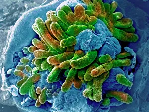
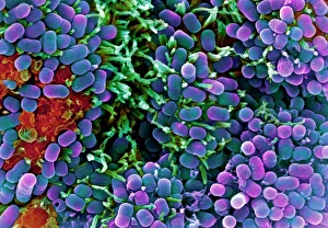
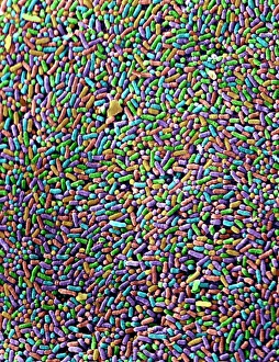
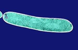
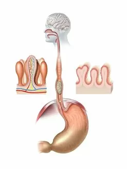
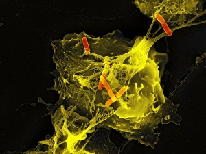
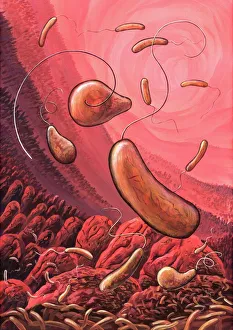
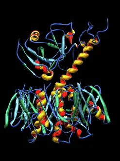
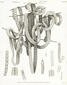
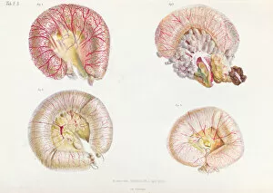
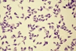
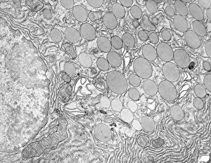
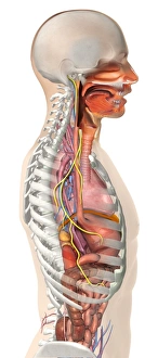
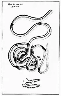
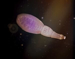
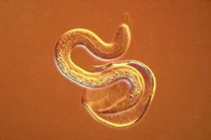
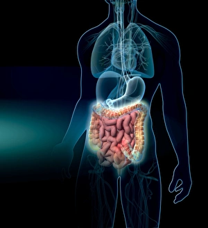
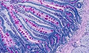
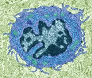
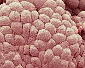
Intestinal health is a complex and fascinating topic that involves various organisms and structures. Through the lens of powerful microscopes, we can explore the intricate world within our digestive system. In one captivating image captured by scanning electron microscopy (SEM), we see the head of a dog tapeworm, reminding us of the diverse parasites that can inhabit our intestines. These microscopic creatures have adapted to survive in this environment, often causing discomfort or illness. Moving deeper into the small intestine, another SEM image reveals its textured surface covered in tiny projections called microvilli. These finger-like structures increase the surface area for nutrient absorption, highlighting their crucial role in digestion. Shifting our focus to bacteria, an SEM snapshot showcases E. Coli bacteria - notorious for causing foodborne illnesses. Their rod-shaped bodies are visible as they navigate through this intestinal landscape. Similarly, Salmonella bacteria make their presence known under SEM; these pathogens can lead to severe gastrointestinal infections if ingested through contaminated food or water. Zooming even closer with transmission electron microscopy (TEM), we observe individual E. coli bacterium with remarkable detail. This high-resolution image allows us to appreciate their cellular structure and understand how they interact within our gut ecosystem. The human digestive system comes alive through artistic representation - a colorful artwork depicting its complexity and interconnectedness. It serves as a reminder of how vital proper digestion is for overall well-being. Exploring further using TEM imagery, we encounter intestinal protozoan parasites such as Cryptosporidium - single-celled organisms that cause diarrheal diseases worldwide. The detailed view provided by TEM helps scientists study these elusive parasites and develop effective treatments against them. Returning to scanning electron microscopy (SEM), we witness yet another dog tapeworm specimen up close – showcasing its segmented body structure designed for survival within an animal host's intestines. Lastly, TEM unveils intestinal microvilli once again but also introduces us to cholera bacteria, depicted in artwork.