Fraud Blocker Skip to main content

Micro Biology Collection (page 5)

Microbiology is a fascinating field that delves into the intricate world of microscopic organisms

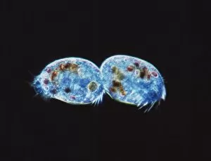
Background imageMicro Biology Collection: Ciliate protozoa dividing

Ciliate protozoa dividing
Ciliate protozoa. Light micrograph of unidentified ciliate protozoa (class Ciliata) undergoing asexual reproduction. These unicellular micro-organisms can multiply by either of two methods

Background imageMicro Biology Collection: Plankton cell wall, SEM

Plankton cell wall, SEM
Plankton cell wall. Coloured scanning electron micrograph (SEM) of the geometrical plates making up the mineralised cell wall of a planktonic alga (Coronosphaera mediterranea)

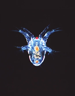
Background imageMicro Biology Collection: Copepod crustacean, light micrograph

Copepod crustacean, light micrograph
Copepod crustacean. Light micrograph of the nauplius larva of a copepod crustacean (subclass Copepoda). This is the first stage in the life cycle of a copepod

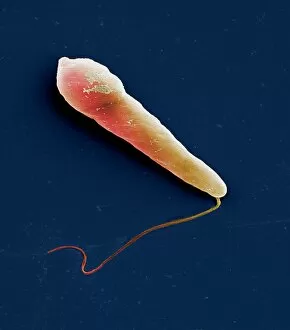
Background imageMicro Biology Collection: Euglena protozoan, SEM

Euglena protozoan, SEM
Euglena protozoan (Euglena sp.), coloured scanning electron micrograph (SEM). This protozoan is described as flagellate because it has a flagellum (thin tail-like structure)

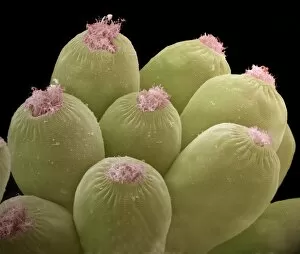
Background imageMicro Biology Collection: Ciliate protozoans, SEM

Ciliate protozoans, SEM
Ciliate protozoans. Coloured scanning electron micrograph (SEM) of sessile (anchored) ciliate protozoan (subclass Peritrichia)

Background imageMicro Biology Collection: Giardia protozoan, TEM

Giardia protozoan, TEM
Giardia protozoan. Coloured transmission electron micrograph (TEM) of a Giardia sp. protozoan. It is a single-celled organism that has two cell nuclei (green)

Background imageMicro Biology Collection: Rotifer worm, light micrograph

Rotifer worm, light micrograph
Rotifer worm. Light micrograph of the rotifer worm Platyas quadricornis. This free-swimming, aquatic micro-organism has a body which is divided into three sections

Background imageMicro Biology Collection: Ciliate protozoa, SEM

Ciliate protozoa, SEM
Ciliate protozoa. Coloured scanning electron micrograph (SEM) of cilliate protozoa (unicellular animals). The ciliates are so named because of the hair-like cilia surrounding their bodies which are

Background imageMicro Biology Collection: Water flea, light micrograph

Water flea, light micrograph
Water flea. Light micrograph of a water flea (suborder Cladocera). The long antennae (upper left and right) of this freshwater crustacean have swimming hairs and serve as rowing organs

Background imageMicro Biology Collection: Flagellate protozoan, SEM

Flagellate protozoan, SEM
Flagellate protozoan, coloured scanning electron micrograph (SEM). This is a protozoan that has a flagellum (thin tail-like structure, lower left)

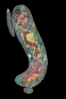
Background imageMicro Biology Collection: Parasitic protozoan, TEM

Parasitic protozoan, TEM
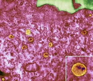
Parasitic protozoan. Coloured transmission electron micrograph (TEM) of a trypanosome protozoan (Trypanosoma sp.). The nucleus (centre), cell membrane (outline)

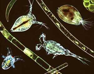
Background imageMicro Biology Collection: Aquatic crustaceans, light micrograph

Aquatic crustaceans, light micrograph
Aquatic crustaceans. Composite image of light micrographs of aquatic planktonic crustaceans with strands of filamentous algae

Background imageMicro Biology Collection: Lychnocanium radiolarian, SEM

Lychnocanium radiolarian, SEM
Lychnocanium radiolarian. Coloured scanning electron micrograph (SEM) of the shell of a Lychnocanium sp. radiolarian. Radiolaria are single-celled protozoans that are found in marine plankton

Background imageMicro Biology Collection: Alga-covered protozoan, SEM

Alga-covered protozoan, SEM
Alga-covered protozoan, coloured scanning electron micrograph (SEM). Algae (blue) are seen on the body of the protozoan, and cilia (green) are used to waft the algae to the protozoans feeding area

Background imageMicro Biology Collection: Paramecium protozoa, SEM

Paramecium protozoa, SEM
Paramecium protozoa (Paramecium sp.), coloured scanning electron micrograph (SEM). These protozoa (two seen) are described as ciliate because many cilia (hair-like structures) cover them

Background imageMicro Biology Collection: Hartmannella amoeba and bacterium

Hartmannella amoeba and bacterium. Computer artwork of a Hartmannella vermiformis amoeba (green) engulfing a Legionella pneumophila bacterium (orange) with a pseudopodium

Background imageMicro Biology Collection: Breslauides ciliate protozoan, SEM

Breslauides ciliate protozoan, SEM
Breslauides ciliate protozoan. Coloured scanning electron micrograph (SEM) of a ciliate protozoan (Breslauides sp.). This tiny single-celled organism is found in water

Background imageMicro Biology Collection: Amphisphaerina radiolarian, SEM

Amphisphaerina radiolarian, SEM
Amphisphaerina radiolarian. Coloured scanning electron micrograph (SEM) of the shell of a Amphisphaerina sp. radiolarian. Radiolaria are single-celled protozoans that are found in marine plankton

Background imageMicro Biology Collection: Acrosphaera radiolarian, SEM

Acrosphaera radiolarian, SEM
Acrosphaera radiolarian. Coloured scanning electron micrograph (SEM) of the shell of a Acrosphaera sp. radiolarian. Radiolaria are single-celled protozoans that are found in marine plankton

Background imageMicro Biology Collection: Green algae colonies

Green algae colonies. Light micrograph of green algae colonies (Volvox sp.). These colonies are made up of approximately 500 individual flagellate cells, arranged in a glycoprotein filled sphere

Background imageMicro Biology Collection: Amoeba, SEM

Amoeba, SEM
Amoeba. Coloured scanning electron micrograph (SEM) of an Amoeba proteus protozoan. This is a freshwater single-celled organism that feeds on bacteria and smaller protozoa

Background imageMicro Biology Collection: Blepharisma ciliate protozoan, SEM

Blepharisma ciliate protozoan, SEM
Blepharisma ciliate protozoan. Coloured scanning electron micrograph (SEM) of a ciliate protozoan (Blepharisma sp.). This tiny single- celled organism is found in freshwater

Background imageMicro Biology Collection: Wardrobe beetle larva, SEM

Wardrobe beetle larva, SEM

Background imageMicro Biology Collection: Fly larva, light micrograph

Fly larva, light micrograph
Fly larva. Light micrograph of the aquatic larva of an unidentified fly (order Diptera). Dark field illumination. Magnification unknown

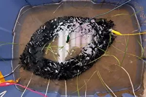
Background imageMicro Biology Collection: Electricity from rice plants

Electricity from rice plants. Electrodes placed in a pot of rice seedlings grown underwater. Anaerobic bacteria, such as Geobacter sp

Background imageMicro Biology Collection: Biofuel bacteria

Biofuel bacteria. Petri dish containing colonies of recombinant (genetically modified) Streptomyces bacteria (red) that may produce cellulase

Background imageMicro Biology Collection: Cell mitosis

Cell mitosis. Confocal fluorescence light micrograph composite showing 6 stages of mitotic cell division. At far left, the cell has completed the first stage of cell division known as interphase in

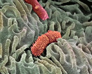
Background imageMicro Biology Collection: Oesophagus lining, SEM

Oesophagus lining, SEM
Oesophagus lining. Coloured scanning electron micrograph (SEM) of the lining (epithelium) of the human oesophagus (gullet, blue) and two bacteria (red)

Background imageMicro Biology Collection: Phagocytosis of a yeast spore, SEM

Phagocytosis of a yeast spore, SEM
Phagocytosis of a yeast spore. Coloured scanning electron micrograph (SEM) of a neutrophil (brown) phagocytosing (engulfing and destroying) a spore from the yeast Candida albicans (orange)

Background imageMicro Biology Collection: Phagocytosis of a fungus spore, SEM

Phagocytosis of a fungus spore, SEM
Phagocytosis of a fungus spore. Coloured scanning electron micrograph (SEM) of a spore from the fungus Aspergillus fumigatus (red) being phagocytosed (engulfed and destroyed)

Background imageMicro Biology Collection: TEM of cluster of P1 bacteriophages

TEM of cluster of P1 bacteriophages

Background imageMicro Biology Collection: Parvovirus particles, TEM

Parvovirus particles, TEM
Parvovirus particles. Coloured transmission electron micrograph (TEM) of virus particles (orange) clustered around a piece of cell membrane (purple, centre)

Background imageMicro Biology Collection: HIV particles in infected cell, TEM

HIV particles in infected cell, TEM
HIV particles. Coloured transmission electron micrograph (TEM) of human immunodeficiency virus (HIV) particles (orange) in a host cell. HIV causes the disease AIDS


All Professionally Made to Order for Quick Shipping

Microbiology is a fascinating field that delves into the intricate world of microscopic organisms. From budding yeast cells to calcareous phytoplankton, these tiny beings hold immense importance in our ecosystem. Take a closer look through the lens of a scanning electron microscope (SEM), and you'll be amazed by their intricate structures. One such example is the avian flu virus, which can cause devastating outbreaks among bird populations. Its unique shape and composition are revealed under SEM, highlighting its potential threat to both animals and humans alike. Diatoms, another group of microorganisms, showcase stunning beauty when observed through SEM. Their delicate silica shells form intricate patterns that resemble works of art. These diatoms play a crucial role in marine ecosystems as primary producers. E. coli bacteria, often associated with foodborne illnesses, reveal their rod-shaped structure under SEM. Understanding their morphology helps scientists develop strategies to combat infections caused by this notorious bacterium. The PSCI2A-00015 embryonic stem cell captured under SEM demonstrates its incredible regenerative capabilities. This versatile cell type holds great promise for future medical advancements and treatments. Salmonella bacteria also come into focus under SEM, displaying their flagella-covered surface responsible for motility and infection transmission. Studying these pathogens aids in developing effective prevention measures against salmonellosis. Intriguingly colored TEM images unveil Yersinia pestis bacteria responsible for causing deadly diseases like bubonic plague throughout history. Unveiling their detailed structure allows researchers to better understand how they function within host organisms. Another captivating diatom species reveals itself through SEM imaging - showcasing nature's artistic side once again. These single-celled algae contribute significantly to global carbon fixation while providing habitats for countless aquatic creatures. Candida fungus captures attention with its filamentous appearance when viewed at high magnification using SEM techniques. This opportunistic pathogen can cause various infections in humans ranging from mild oral thrush to life-threatening systemic infections.