Fraud Blocker Skip to main content

Micro Biology Collection (page 2)

Microbiology is a fascinating field that delves into the intricate world of microscopic organisms

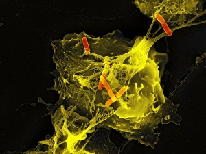
Background imageMicro Biology Collection: Neutrophil cell trapping bacteria, SEM

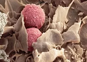
Neutrophil cell trapping bacteria, SEM
Neutrophil cell trapping bacteria. Coloured scanning electron micrograph (SEM) of bacteria (rod-shaped) being trapped by a neutrophil cell

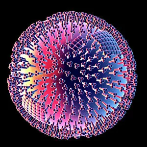
Background imageMicro Biology Collection: Influenza virus, computer artwork

Influenza virus, computer artwork
Influenza virus. Computer artwork of an influenza (flu) virus. The virus consists of a core of RNA (ribonucleic acid) genetic material surrounded by a protein coat

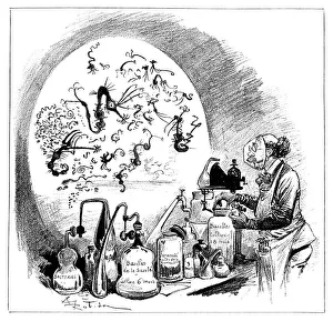
Background imageMicro Biology Collection: Microbiology caricature, 19th century

Microbiology caricature, 19th century
Microbiology caricature. The researcher is handling jars labelled as bacterial cultures, while the magnified view at left shows a caricatured appearance of microbes as seen under a microscope

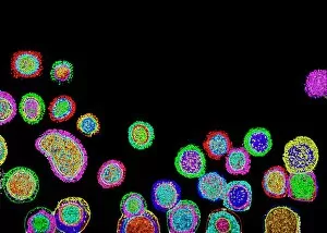
Background imageMicro Biology Collection: Influenza virus particles, TEM

Influenza virus particles, TEM
Influenza virus particles, coloured transmission electron micrograph (TEM). This virus is the cause of the respiratory disease influenza (flu) in humans and other animals

Background imageMicro Biology Collection: Hepatitis C viruses, TEM

Hepatitis C viruses, TEM
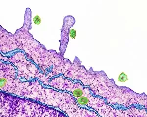
Hepatitis C viruses. Coloured transmission electron micrograph (TEM) of hepatitis C virus particles (green) infecting cultured liver cells (purple)

Background imageMicro Biology Collection: Cell division, fluorescent micrograph

Cell division, fluorescent micrograph
Cell division. Immunofluorescent light micrograph of a human epithelial cell (centre) during the interphase stage of mitosis

Background imageMicro Biology Collection: Louis Pasteur, French microbiologist

Louis Pasteur, French microbiologist
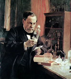
Louis Pasteur (1822-1895), French microbiologist and chemist, in his laboratory. Louis Pasteur proved the germ theory of disease, which holds that germs attack the body from the outside

Background imageMicro Biology Collection: Dinoflagellate plankton, SEM

Dinoflagellate plankton, SEM
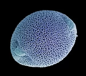
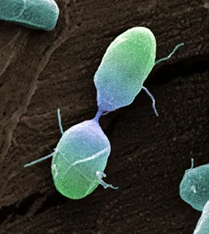
Dinoflagellate plankton. Coloured scanning electron micrograph (SEM) of the calcareous (calcium-based) test (shell) of a dinoflagellate Calciodinellum sp

Background imageMicro Biology Collection: Pseudomonas aeruginosa bacteria, SEM

Pseudomonas aeruginosa bacteria, SEM
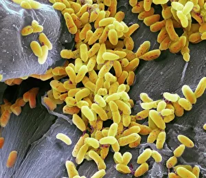
Pseudomonas aeruginosa bacteria, coloured scanning electron micrograph (SEM). These Gram-negative rod-shaped bacteria are found in soil and water, and as normal flora in the human intestine

Background imageMicro Biology Collection: Immunofluorescent LM of fibroblast cell nuclei

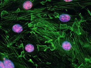
Immunofluorescent LM of fibroblast cell nuclei
Cell nuclei. Immunofluorescent light micrograph of cultured fibroblast cells showing nuclei with " nucleolar necklaces"

Background imageMicro Biology Collection: Adenovirus, artwork

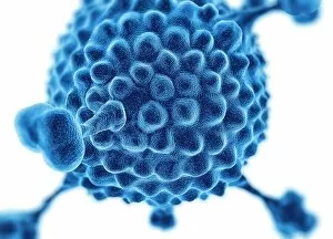
Adenovirus, artwork
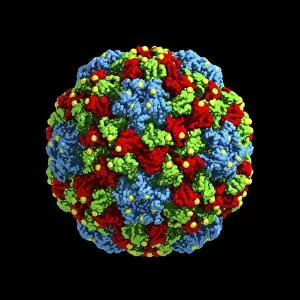
Adenovirus. Computer artwork of an adenovirus, showing the surface structure of the viruss outer protein coat (capsid). Adenoviruses are known to infect humans

Background imageMicro Biology Collection: TEM of Lambda bacteriophages

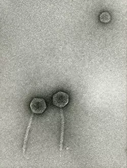
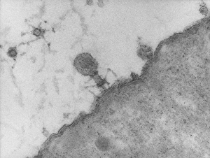
TEM of Lambda bacteriophages

Background imageMicro Biology Collection: Salmonella culture

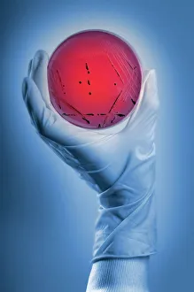
Salmonella culture
MODEL RELEASED. Salmonella culture. Microbiologist holding a petri dish containing a culture of Salmonella sp. bacteria. These bacteria are the cause of salmonellosis (food poisoning) in humans

Background imageMicro Biology Collection: Mouse malaria parasite, SEM

Mouse malaria parasite, SEM
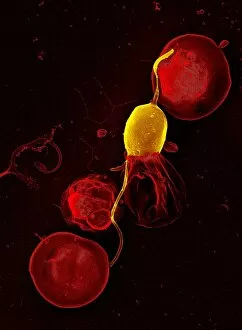
Mouse malaria parasite. Coloured scanning electron micrograph (SEM) of a Plasmodium berghei protozoan (yellow) and red blood cells. P. berghei is the parasite that causes malaria in mice

Background imageMicro Biology Collection: Rift Valley fever virus, TEM

Rift Valley fever virus, TEM

Background imageMicro Biology Collection: Vesicular stomatitis virus, TEM

Vesicular stomatitis virus, TEM
Vesicular stomatitis virus. Coloured transmission electron micrograph (TEM) of vesicular stomatitis virus (VSV) particles (blue) budding from a host cell (red)

Background imageMicro Biology Collection: Paramyxovirus particles, TEM

Paramyxovirus particles, TEM
Sendai virus. Coloured transmission electron micrograph (TEM) of Sendai virus particles (virions, orange). The protein coat (capsid) of one of the particles has split

Background imageMicro Biology Collection: Self-replicating nanobots, artwork

Self-replicating nanobots, artwork
Self-replicating nanobots, conceptual computer artwork

Background imageMicro Biology Collection: Mycobacterium chelonae bacteria, SEM

Mycobacterium chelonae bacteria, SEM
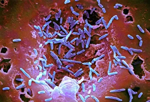
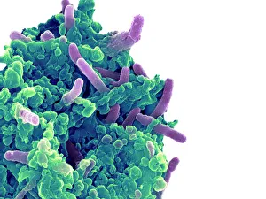
Mycobacterium chelonae bacteria, coloured scanning electron micrograph (SEM). These Gram-positive rod-shaped bacteria are found in soil and water

Background imageMicro Biology Collection: Frozen sperm, conceptual image

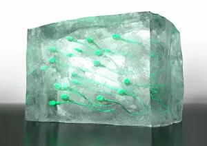
Frozen sperm, conceptual image. Computer artwork of sperm cells frozen inside a block of ice, representing the long term preservation of donated sperm to be used in fertility treatments

Background imageMicro Biology Collection: E. coli bacterium, TEM

E. coli bacterium, TEM
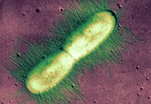
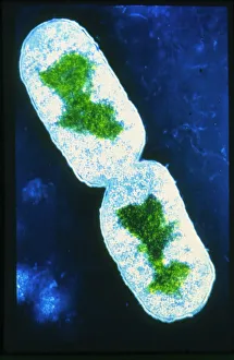
E. coli bacterium. Coloured transmission electron micrograph (TEM) of an Escherichia coli bacterium in the early stages of binary fission, the process by which the bacterium divides

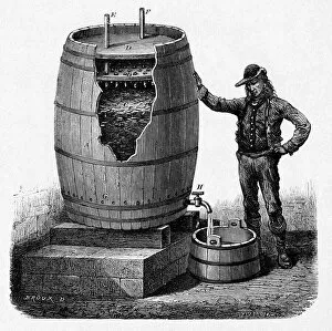
Background imageMicro Biology Collection: Vinegar production, 19th century

Vinegar production, 19th century cutaway artwork. This is a trickling method developed in 1823 by the German chemist Schutzenbach. The barrel is divided into three sections

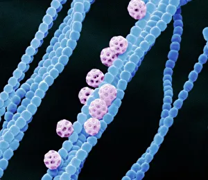
Background imageMicro Biology Collection: Cyanobacteria, SEM

Cyanobacteria, SEM
Cyanobacteria. Coloured scanning electron micrograph (SEM) of cyanobacteria (formally known as blue-green algae, blue). Several spores (pink) are also seen

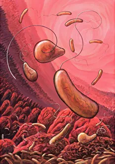
Background imageMicro Biology Collection: Cholera bacteria, artwork

Cholera bacteria, artwork
Cholera bacteria. Artwork of Vibrio cholerae bacteria in the small intestine. These Gram-negative rod-shaped bacteria have a single polar flagellum (tail-like structure)

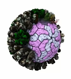
Background imageMicro Biology Collection: Rotavirus particle, artwork

Rotavirus particle, artwork
Rotavirus particle. Cut-away artwork showing the structure of the rotavirus icosahedral capsid (protein coat). The capsid, which encloses the RNA (ribonucleic acid) genome

Background imageMicro Biology Collection: E. coli 0157: H7 bacteria

E. coli 0157: H7 bacteria
E. coli 0157:H7 bacteria. Coloured scanning electron micrograph (SEM) of Escherichia coli 0157:H7 bacteria, cause of foodborne illness

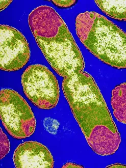
Background imageMicro Biology Collection: Salmonella bacterium dividing, SEM

Salmonella bacterium dividing, SEM
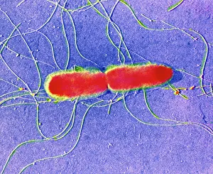
Salmonella bacterium dividing. Coloured scanning electron micrograph (SEM) of of a Salmonella bacterium dividing. The two new daughter cells are seen at upper right and lower left

Background imageMicro Biology Collection: Bacteria infecting a macrophage, SEM

Bacteria infecting a macrophage, SEM
Bacteria infecting a macrophage. Coloured scanning electron micrograph (SEM) of Mycobacterium tuberculosis bacteria (purple) infecting a macrophage white blood cell

Background imageMicro Biology Collection: False-colour TEM of Salmonella typhi

False-colour TEM of Salmonella typhi

Background imageMicro Biology Collection: Cholera toxin, molecular model

Cholera toxin, molecular model
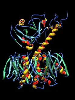
Cholera toxin. Molecular model of the secondary structure of cholera enterotoxin (intestinal toxin). The molecule consists of two subunits, A (top) and B (bottom)

Background imageMicro Biology Collection: False-colour TEM of bacterium E. Coli

False-colour TEM of bacterium E. Coli
False colour transmission electron micrograph of the bacterium Escherichia coli, a normal member of human intestinal flora

Background imageMicro Biology Collection: E. coli bacterium dividing

E. coli bacterium dividing

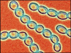
Background imageMicro Biology Collection: Chains of Streptococcus pyogenes bacteria

Chains of Streptococcus pyogenes bacteria

Background imageMicro Biology Collection: International biohazard symbol

International biohazard symbol warning of a potential biological hazard. This symbol is used worldwide in laboratories and on containers where biologically active agents are present

Background imageMicro Biology Collection: Phagocytosis of fungus spores, SEM

Phagocytosis of fungus spores, SEM
Phagocytosis of fungus spores. Coloured scanning electron micrograph (SEM) of spores from the fungus Aspergillus fumigatus (red) being phagocytosed (engulfed and destroyed) by a dendritic cell (brown)

Background imageMicro Biology Collection: Anton van Leeuwenhoek, caricature

Anton van Leeuwenhoek, caricature
Anton van Leeuwenhoek. Caricature of the Dutch microbiologist Anton van Leeuwenhoek (1632-1723), looking through a magnifying glass to represent his discoveries in microbiology

Background imageMicro Biology Collection: Giardia lamblia protozoa, micrograph

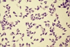
Giardia lamblia protozoa, micrograph
Giardia lamblia protozoa, light micrograph. These single-celled parasitic protozoa infect the intestinal tract and are most common in tropical regions

Background imageMicro Biology Collection: Antibiotic action, historical image

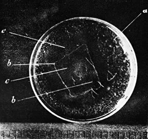
Antibiotic action, historical image
Antibiotic action. Historical image taken by the German pathologist Karl Dohle (1855-1928) in 1889, showing the action of an antibiotic compound on Bacillus anthracis (anthrax)

Background imageMicro Biology Collection: Botulism bacteria

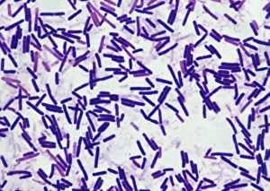
Botulism bacteria. Light micrograph of botulism bacteria (Clostridium botulinum). These bacteria occur naturally in soil. They produce botulinum toxin

Background imageMicro Biology Collection: Yeast cells, SEM

Yeast cells, SEM
Yeast cells. Coloured scanning electron micrograph (SEM) of cells of bakers yeast (Saccharomyces cerevisiae) from part of a dried, commercial yeast pellet

Background imageMicro Biology Collection: Paramecium bursaria protozoan, micrograph

Paramecium bursaria protozoan, micrograph
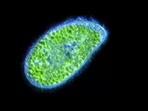
Paramecium bursaria protozoan, light micrograph. This ciliate protozoan inhabits freshwater, where it feeds mainly on bacteria

Background imageMicro Biology Collection: Creation of oil using designer microbes

Creation of oil using designer microbes. Conceptual computer artwork depicting an oil well pump in a petri dish, representing the use of artificially created micro-organisms to produce oil

Background imageMicro Biology Collection: TEM of T4 bacteriophage infecting E. coli

TEM of T4 bacteriophage infecting E. coli
Transmission electron micrograph of bacteriophage T4 (virus infecting bacteria), 60 minutes after the injection of its viral DNA into the bacterium Escherichia coli

Background imageMicro Biology Collection: Cucumber mosaic virus, computer model

Cucumber mosaic virus, computer model
Cucumber mosaic virus (CMV), computer model. This image was created using molecular modelling software and data from X-ray crystallography

Background imageMicro Biology Collection: Creation of artificial life, artwork

Creation of artificial life, artwork
Creation of artificial life. Conceptual computer artwork depicting the creation of a new life-form in a petri dish. Geneticists are working on methods for combining artificially replicated genes into

Background imageMicro Biology Collection: Anthrax antibiotics research

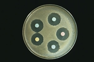
Anthrax antibiotics research. Antibiotic drugs (discs) being tested on anthrax bacteria (Bacillus anthracis) colonies being cultured on an agar growth medium in a petri dish

Background imageMicro Biology Collection: HIV viruses, TEM

HIV viruses, TEM
HIV viruses. Coloured transmission electron micrograph (TEM) of human immunodeficiency virus (HIV) particles. These particles are docking at a T-lymphocyte (white blood cell) just before infection

Background imageMicro Biology Collection: Louis Pasteur, French chemist

Louis Pasteur, French chemist


All Professionally Made to Order for Quick Shipping

Microbiology is a fascinating field that delves into the intricate world of microscopic organisms. From budding yeast cells to calcareous phytoplankton, these tiny beings hold immense importance in our ecosystem. Take a closer look through the lens of a scanning electron microscope (SEM), and you'll be amazed by their intricate structures. One such example is the avian flu virus, which can cause devastating outbreaks among bird populations. Its unique shape and composition are revealed under SEM, highlighting its potential threat to both animals and humans alike. Diatoms, another group of microorganisms, showcase stunning beauty when observed through SEM. Their delicate silica shells form intricate patterns that resemble works of art. These diatoms play a crucial role in marine ecosystems as primary producers. E. coli bacteria, often associated with foodborne illnesses, reveal their rod-shaped structure under SEM. Understanding their morphology helps scientists develop strategies to combat infections caused by this notorious bacterium. The PSCI2A-00015 embryonic stem cell captured under SEM demonstrates its incredible regenerative capabilities. This versatile cell type holds great promise for future medical advancements and treatments. Salmonella bacteria also come into focus under SEM, displaying their flagella-covered surface responsible for motility and infection transmission. Studying these pathogens aids in developing effective prevention measures against salmonellosis. Intriguingly colored TEM images unveil Yersinia pestis bacteria responsible for causing deadly diseases like bubonic plague throughout history. Unveiling their detailed structure allows researchers to better understand how they function within host organisms. Another captivating diatom species reveals itself through SEM imaging - showcasing nature's artistic side once again. These single-celled algae contribute significantly to global carbon fixation while providing habitats for countless aquatic creatures. Candida fungus captures attention with its filamentous appearance when viewed at high magnification using SEM techniques. This opportunistic pathogen can cause various infections in humans ranging from mild oral thrush to life-threatening systemic infections.