
Parasite Collection
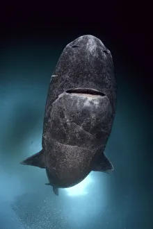
"Unseen Intruders: The Fascinating World of Parasites" A Greenland shark (Somniosus microcephalus) with a parasitic copepod (Ommatokoita elongata) clinging to its skin


All Professionally Made to Order for Quick Shipping
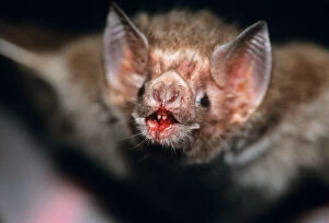
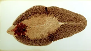
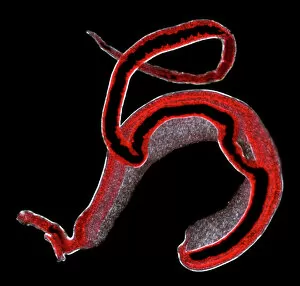
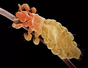
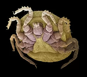
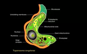
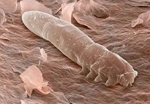
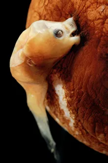
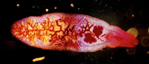
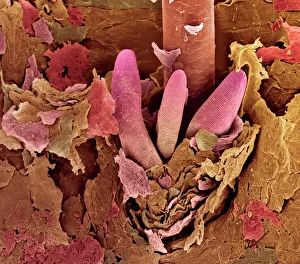
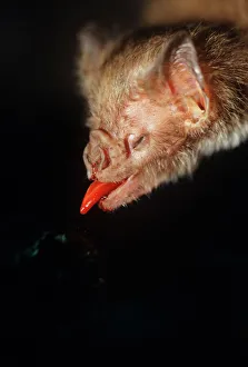
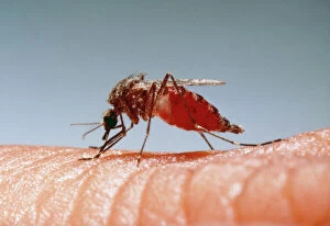
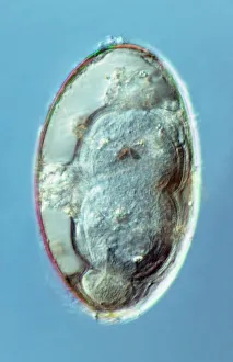
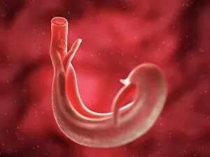
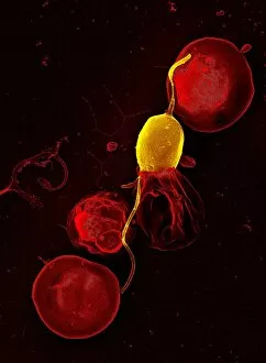
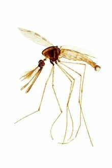
"Unseen Intruders: The Fascinating World of Parasites" A Greenland shark (Somniosus microcephalus) with a parasitic copepod (Ommatokoita elongata) clinging to its skin, showcasing the intricate relationship between host and parasite. Get up close and personal with the Common Vampire Bat's face after feeding in Sao Paulo, Brazil - a reminder of nature's bloodsucking specialists. Delve into the microscopic world as we explore a liver fluke under slide preparation, revealing the complex anatomy of this notorious internal parasite. Unraveling the secrets of Schistosoma spp. , blood flukes that cause debilitating diseases in humans, through detailed examination and research. Witness the astonishing head structure of a dog tapeworm under SEM, highlighting its ability to attach itself firmly within its host's intestines. Discover an incredibly detailed SEM image capturing every tiny feature of a head louse - an unwelcome guest causing discomfort worldwide. Zooming in on a sheep tick using SEM reveals its formidable mouthparts designed for piercing and sucking blood from unsuspecting hosts. Observe nature's macabre beauty as a parasitic wasp lays eggs inside hoverfly larvae, showcasing their cunning reproductive strategy right before our eyes in the UK countryside. Immerse yourself in stunning artwork depicting Trypanosome protozoan parasites responsible for deadly sleeping sickness across Africa - reminding us how art can convey scientific knowledge too. Explore Plasmodium sp. , malarial parasites that wreak havoc on millions worldwide each year, through captivating visuals that shed light on their life cycle and impact on human health. Peer into the hidden world of eyelash mites using SEM imagery; these tiny creatures inhabit our lashes without us even realizing it.