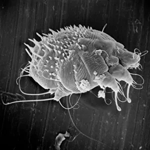
Plasmodium sp. malarial parasite

![]()
Wall Art and Photo Gifts from Mary Evans Picture Library
Plasmodium sp. malarial parasite
Scanning electron microscope image of a malarial protozoal parasite. The parasite requires the anopheles mosquito to complete its life cycle
Mary Evans Picture Library makes available wonderful images created for people to enjoy over the centuries
Media ID 8611790
© Mary Evans Picture Library 2015 - https://copyrighthub.org/s0/hub1/creation/maryevans/MaryEvansPictureID/10711607
Cell Disease Electron Electron Micrograph Eukaryote Eukaryotic Malaria Micrograph Microscope Microscope Image Parasite Protist Protista Protozoa Protozoan Scanning Scanning Electron Micrograph Scanning Electron Microscope Scanning Electron Microscope Image Sem Image Plasmodium
EDITORS COMMENTS
1. Title: A Microscopic Marvel: The Plasmodium Species Malarial Parasite. This Scanning Electron Microscope (SEM) image showcases the intricate structure of a Plasmodium sp. malarial parasite. Malaria is a mosquito-borne infectious disease that affects millions of people worldwide, causing symptoms ranging from fever, headache, and muscle pain to severe complications such as organ failure and death. The Plasmodium parasite is a unicellular eukaryote, belonging to the phylum Apicomplexa and the class Sporozoasida. It requires the Anopheles mosquito as its vector to complete its complex life cycle. In the human host, the parasite invades red blood cells, causing the characteristic symptoms of malaria. In this image, the parasite's surface is revealed in stunning detail. The intricate, undulating membrane of the parasitic cell is adorned with tiny projections, or micronemes, which are believed to play a role in the initial attachment of the parasite to the red blood cell. The parasite's unique morphology is a testament to the intricacy and diversity of the microscopic world. The Plasmodium parasite is a master of adaptation, with five distinct species capable of causing human malaria: Plasmodium falciparum, Plasmodium vivax, Plasmodium ovale, Plasmodium malariae, and Plasmodium knowlesi. Each species exhibits subtle differences in its life cycle and disease manifestations, making the study of these parasites crucial for the development of effective malaria diagnostics, treatments, and vaccines. This SEM image offers a glimpse into the microscopic world of the Plasmodium parasite, revealing its intricate structure and shedding light on the complex biology of this fascinating and deadly organism.
MADE IN AUSTRALIA
Safe Shipping with 30 Day Money Back Guarantee
FREE PERSONALISATION*
We are proud to offer a range of customisation features including Personalised Captions, Color Filters and Picture Zoom Tools
SECURE PAYMENTS
We happily accept a wide range of payment options so you can pay for the things you need in the way that is most convenient for you
* Options may vary by product and licensing agreement. Zoomed Pictures can be adjusted in the Cart.