
Bristle Collection (page 2)
"Bristle: Nature's Intricate Design" From the delicate bristles on a moth antenna to the meticulous brushing of teeth, a fascinating element found in various forms


All Professionally Made to Order for Quick Shipping
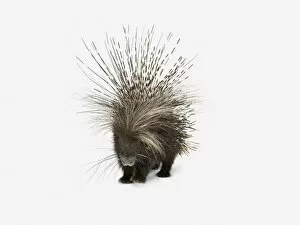
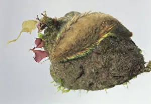
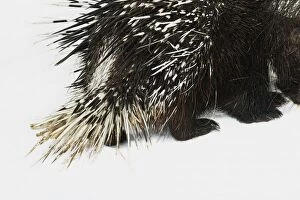
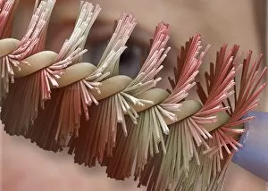
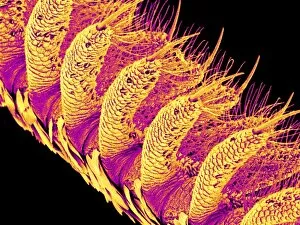
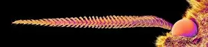
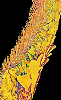
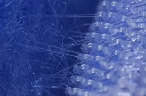
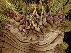
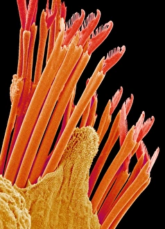
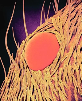
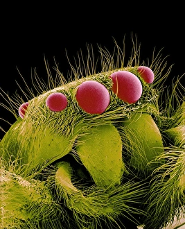
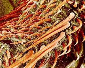
"Bristle: Nature's Intricate Design" From the delicate bristles on a moth antenna to the meticulous brushing of teeth, a fascinating element found in various forms. Under the watchful eye of an SEM (Scanning Electron Microscope), these tiny structures reveal their intricate beauty. In the botanical world, changeable velvetberry, scientifically known as Stachytarpheta mutabilis, showcases its vibrant petals adorned with soft bristles. Turn to page 226 and discover the enchanting Fringed Helmet Orchid (Corysanthes fimbriata Br / aFringed) with its velvety texture and exquisite bristled appearance. Venturing into fern territory, we encounter Trichomanes elegans - commonly referred to as Bristle Fern. Its slender fronds are adorned with fine hairs that add an ethereal touch to any landscape. Another member of this family is Trichomanes botryoides; its unique structure captivates all who stumble upon it. Diving beneath the waves, we delve into marine life where bristle worms or polychaetes reign supreme. These segmented creatures display a remarkable array of colorful bristles along their bodies - nature's own work of art. Sea mice or Bristle worms like Arenicola marina and Clymene amphistoma leave trails behind them as they navigate through sandy ocean floors. Amphitrite ventilabrum emerges from undersea depths; this sea worm boasts magnificent feathery appendages resembling delicate brushes made up of countless tiny bristles. The sight is truly mesmerizing. Returning to land once more, we find ourselves amidst various species of Trichomanes ferns - each showcasing their distinct charm through elegant fronds embellished with minute hair-like projections. Bristles exist in myriad forms across different realms - from insect antennae to plant petals and marine organisms' bodies.