
Calcified Collection
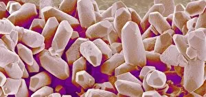
"Exploring the Fascinating World Wonders" Delving into the depths of bone structure, a light micrograph reveals the intricate beauty of compact bone


All Professionally Made to Order for Quick Shipping
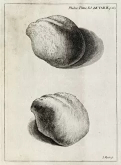
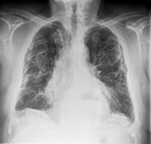
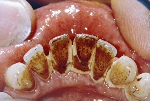
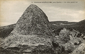
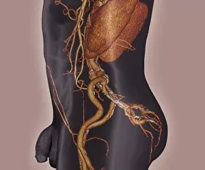
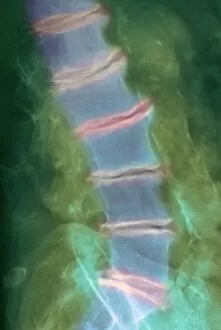
"Exploring the Fascinating World Wonders" Delving into the depths of bone structure, a light micrograph reveals the intricate beauty of compact bone, showcasing its strength and resilience. Travel back in time to the 18th century when kidney stones were first discovered as calcified formations within our bodies, reminding us of the marvels hidden within. Pamukkale's hot springs offer a breathtaking sight with their stunning calcified deposits, creating cascading terraces that seem like nature's own masterpiece. Winter transforms Yellowstone National Park into a magical wonderland where even trees become calcified sculptures at Tangled Creek, standing tall against the snowy backdrop. Witnessing nature's artistry in Algeria's Hammam Maskhoutine, an ancient dried geyser cone stands as a testament to time and showcases its unique calcification process. Dental hygiene takes center stage as dental plaque and tartar remind us of the importance of regular care to prevent these unwanted calcifications from forming on our teeth. Returning once again to Yellowstone National Park's Tangled Creek in winter, we are captivated by the ethereal beauty of frozen trees transformed into magnificent calcified structures. Asbestosis leaves its mark on X-ray images like C017/7143, revealing how harmful mineral fibers can lead to lung tissue scarring and subsequent calcium deposition over time. The ovary becomes an unexpected canvas for calcification as cysts form within it, highlighting both medical challenges and fascinating biological phenomena occurring inside our bodies. A rough irregular shell encloses an oyster tightly while displaying layered segments and ridges—a captivating glimpse into nature’s way of protecting delicate creatures through natural calcification processes. Bacon-wrapped oyster flesh delicately draped across shells secured with toothpicks creates an appetizing presentation atop coarse salt—an exquisite blend between culinary art and the calcified shells of these ocean delicacies.