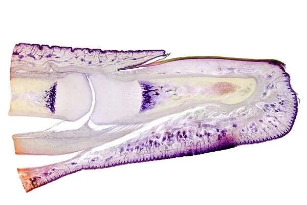
Human finger, longitudinal section
![]()

Wall Art and Photo Gifts from Science Photo Library
Human finger, longitudinal section
Human finger. Light micrograph of a longitudinal section through a finger of a human infant. This shows the bones inside the finger (here, the 1st and 2nd phalanges). Each phalanx has outer compact bone (yellow) and an inner marrow. The cartilaginous ends of the bone are undergoing growth (ossification) and the bluish-purple layer is undergoing calcification. The skin consists of an outer epidermis (purple) and a lower dermis in which sweat glands and ducts (purple dots) are seen. The nail (upper right) is made up of a hard protein (keratin), which is produced by the nail root (upper centre). Magnification: x14 when printed at 10 centimetres across
Science Photo Library features Science and Medical images including photos and illustrations
Media ID 6278855
© DR KEITH WHEELER/SCIENCE PHOTO LIBRARY
Bones Calcification Calcified Cartilage Child Compact Bone Cuticle Dermis Epidermis Finger Glands Growth Histological Histology Infant Joint Joints Keratin Keratinised Knuckle Knuckles Longitudinal Marrow Nail Ossification Phalanges Phalanx Skin Sweat Gland Tissue Light Micrograph Light Microscope Section Sectioned
EDITORS COMMENTS
This print from Science Photo Library showcases a longitudinal section of a human finger, specifically that of an infant. Through the lens of a light microscope, we are granted a glimpse into the intricate anatomy and growth processes within this tiny digit. The image reveals the bones nestled inside the finger - in this case, the first and second phalanges. Each phalanx exhibits both outer compact bone, depicted in yellow hues, and inner marrow. Notably, we witness the cartilaginous ends of these bones undergoing ossification or growth while a bluish-purple layer signifies calcification. Beyond its skeletal structure lies the skin comprising two layers: an outer epidermis shown in purple tones and a lower dermis housing sweat glands and ducts represented by purple dots. The upper right corner features a nail composed of keratin, produced by the nail root situated at the upper center. With magnification set at x14 when printed at 10 centimeters across, this remarkable photograph provides us with valuable insights into various biological aspects such as tissue composition, joint formation, normal development patterns in infants' fingers. It serves as an educational tool for understanding human anatomy while evoking curiosity about our own bodies' complexity.
MADE IN AUSTRALIA
Safe Shipping with 30 Day Money Back Guarantee
FREE PERSONALISATION*
We are proud to offer a range of customisation features including Personalised Captions, Color Filters and Picture Zoom Tools
SECURE PAYMENTS
We happily accept a wide range of payment options so you can pay for the things you need in the way that is most convenient for you
* Options may vary by product and licensing agreement. Zoomed Pictures can be adjusted in the Cart.

