
Keratinised Collection
"Exploring the Fascinating World Surfaces: From Skin to Hair" Delving into the intricate details surfaces


All Professionally Made to Order for Quick Shipping
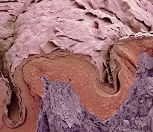
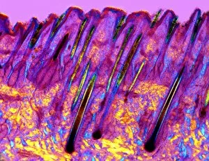
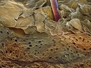
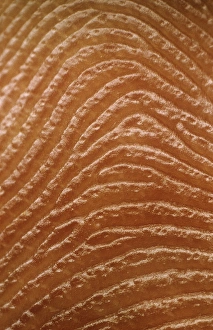
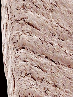
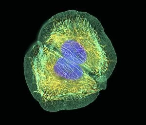
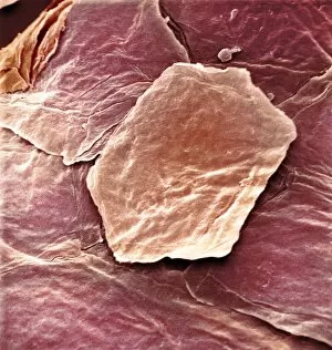
"Exploring the Fascinating World Surfaces: From Skin to Hair" Delving into the intricate details surfaces, we examine the skin surface under a scanning electron microscope (SEM), revealing its remarkable texture and composition. Zooming in further, we explore human hair strands using SEM, uncovering their unique structure and the role of keratin in providing strength and protection. Continuing our journey through microscopic wonders, another SEM image showcases the complexity of human hair at an even higher magnification level, highlighting its incredible resilience and beauty. Shifting gears to foetal skin development, SEM images C016/9094 and C016/9093 offer a glimpse into the early stages of keratinisation, shedding light on how this process shapes our skin's barrier function. An artistic representation (artwork C016/7541) takes us beneath the surface to unveil the intricate layers that make up our skin structure - a testament to nature's flawless design. Examining finger skin through SEM reveals its unique patterned ridges and valleys that aid in grip and tactile sensitivity – a true marvel of evolution. Another captivating SEM image captures finger skin from a different perspective, showcasing its distinct cellular arrangement with precision and clarity. A beautifully illustrated artwork guides us through the complex anatomy of human skin - an organ that not only protects but also communicates with our environment in countless ways. Venturing beyond traditional boundaries, we delve into tongue tissue using polarised light micrograph C015/7105; here too lies evidence of keratinisation playing a vital role in maintaining oral health. Returning to familiar territory yet still awe-inspiring, an SEM image unveils more secrets hidden within our largest organ - revealing both delicate structures like pores as well as robust barriers against external threats.