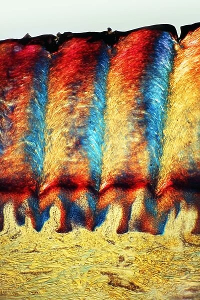
Heel skin tissue, light micrograph
![]()

Wall Art and Photo Gifts from Science Photo Library
Heel skin tissue, light micrograph
Heel skin tissue. Polarised light micrograph of a transverse section through skin from the heel of a human foot. The sole of the foot has to withstand the weight of the body and friction when the foot pushes against the substratum. To cope with this, the outer epidermis forms deep layers of dead keratinised cells (red and blue) which are replaced from the meristematic Malpighian layer as they get worn away. The dermis (yellow and green) consists of collagenous elastic fibres. Magnification: x103 when printed at 10 centimetres high
Science Photo Library features Science and Medical images including photos and illustrations
Media ID 6278345
© DR KEITH WHEELER/SCIENCE PHOTO LIBRARY
Collagen Cross Section Dead Deep Dermal Dermatological Dermatology Dermis Epidermal Epidermis Foot Histological Histology Keratinised Polarised Polarized Skin Sole Surface Thick Tissue Tough Light Micrograph Light Microscope Section Sectioned
EDITORS COMMENTS
This print showcases the intricate structure of heel skin tissue. Taken under polarised light, this transverse section reveals the remarkable adaptations that enable our feet to withstand the weight of our bodies and friction against various surfaces. The outer epidermis, depicted in vibrant shades of red and blue, forms deep layers of dead keratinised cells. These resilient cells act as a protective barrier for the foot's sole, constantly replenishing themselves from the meristematic Malpighian layer as they wear away over time. Beneath the epidermis lies the dermis, illustrated in hues of yellow and green. Composed of collagenous elastic fibers, this layer provides essential support and flexibility to ensure proper functioning of the foot. The interplay between these two layers is crucial for maintaining healthy heel skin tissue. At a magnification level of x103 when printed at 10 centimeters high, this micrograph allows us to appreciate the intricacies within our own bodies on a microscopic scale. It serves as a reminder that even seemingly ordinary parts like our heels possess extraordinary biological complexity. Whether you're fascinated by biology or simply intrigued by human anatomy, this print offers an intriguing glimpse into one aspect of our body's incredible resilience and adaptability - all captured beautifully by Science Photo Library.
MADE IN AUSTRALIA
Safe Shipping with 30 Day Money Back Guarantee
FREE PERSONALISATION*
We are proud to offer a range of customisation features including Personalised Captions, Color Filters and Picture Zoom Tools
SECURE PAYMENTS
We happily accept a wide range of payment options so you can pay for the things you need in the way that is most convenient for you
* Options may vary by product and licensing agreement. Zoomed Pictures can be adjusted in the Cart.

