
Polarized Collection
"Polarized: Exploring the Intricate World Under the Microscope and Beyond" Delving into the depths of compact bone structure


All Professionally Made to Order for Quick Shipping
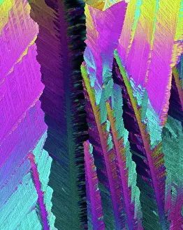
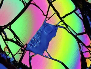
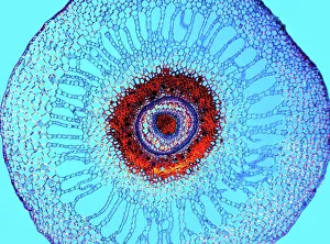
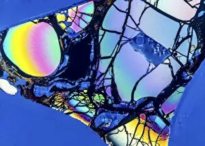
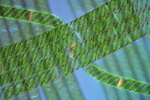
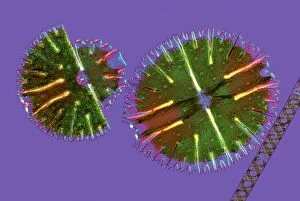
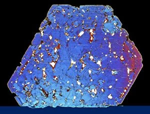
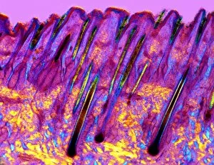
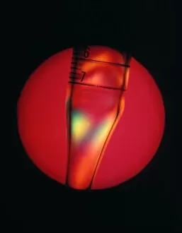
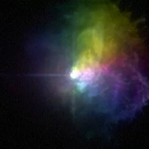
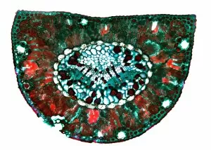
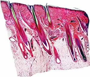
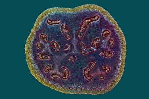
"Polarized: Exploring the Intricate World Under the Microscope and Beyond" Delving into the depths of compact bone structure, a light micrograph reveals its intricate network, showcasing nature's remarkable engineering. Caffeine crystals come to life under the lens, their vibrant patterns resembling a mesmerizing work of art in this captivating light micrograph. Meteorite Jepara takes on an otherworldly appearance when examined as a thin section under a light microscope, transporting us to distant cosmic realms. The water fern rhizome unveils its hidden beauty through a stunning light micrograph, showcasing delicate tendrils that support life beneath aquatic surfaces. A pine tree stem unravels its secrets in vivid detail under the microscope's gaze, displaying concentric rings and textures that tell tales of resilience and growth. The elm stem comes alive with intricate patterns and cellular structures when observed through a light micrograph, revealing nature's elegant craftsmanship. Magnified by the lens, the dog rose stem showcases its thorny armor and vascular system in exquisite detail—a testament to survival strategies honed over time. In Malaysia's Mabul Island paradise, witness an awe-inspiring scene as a Hawksbill Turtle gracefully swims amidst vibrant reefs—an enchanting display of marine biodiversity. Gout crystals take center stage as they are magnified under intense scrutiny—sharp-edged formations hint at painful realities faced by those afflicted with this condition. Whale bone tissue offers glimpses into these majestic creatures' lives; illuminated by microscopic examination, it tells stories of strength and adaptation within their skeletal framework. Black Striped Salema fish from Galapagos Islands captivate our attention with their distinctive markings—nature's brushstrokes creating living artwork beneath ocean waves. A polarising filter transforms ordinary scenes into extraordinary masterpieces; artwork C017 / 7125 showcases the power of light manipulation, revealing hidden dimensions.