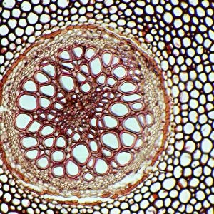
Bracken fern rhizome, light micrograph

![]()
Wall Art and Photo Gifts from Science Photo Library
Bracken fern rhizome, light micrograph
Bracken fern rhizome. Polarised light micrograph of a cross-section through a rhizome from a bracken fern (Pteris aquilinum). The outer layer (yellow-green) consists of supporting fibres. Inside the stem are several meristeles, areas of vascular tissue. The outer layer of each meristele is an endoderm of single cells (dark brown). Below these are the pericycle and phloem cells (lighter brown). At the centre of the meristeles is a lattice of xylem tissue. The phloem transports nutrients, while the xylem transports water and minerals. Magnification: x20 when printed at 10 centimetres across
Science Photo Library features Science and Medical images including photos and illustrations
Media ID 6333551
© DR KEITH WHEELER/SCIENCE PHOTO LIBRARY
Cellular Cross Section Endoderm Fern Nutrient Transport Pericycle Phloem Polarised Light Micrograph Polarised Light Microscopy Polarized Rhizome Supporting Fibres Tissue Transverse Vascular Bundle Vascular Bundles Water Transport Xylem Cells Light Micrograph Light Microscope Section Sectioned
EDITORS COMMENTS
This print showcases the intricate beauty of a Bracken fern rhizome, captured under polarised light microscopy. The image reveals a cross-section through the rhizome, providing a glimpse into its complex cellular structure and vascular system. The outer layer of the rhizome is composed of vibrant yellow-green supporting fibres, which provide structural integrity to the plant. Inside this protective layer, several meristeles are visible as areas of specialized vascular tissue. Each meristele is surrounded by an endoderm consisting of dark brown single cells. Beneath the endoderm lie layers of pericycle and phloem cells in lighter shades of brown. These components play crucial roles in nutrient transport within the plant. At the center of each meristele lies a lattice-like arrangement of xylem tissue responsible for water and mineral transportation. With a magnification level that allows for printing at 10 centimeters across while maintaining clarity, this extraordinary photograph enables us to appreciate nature's complexity on a microscopic scale. It serves as a testament to the wonders found within even seemingly ordinary plants like bracken ferns. This visually stunning image not only appeals to botany enthusiasts but also provides valuable insights into biological research and education fields such as cell biology, botanical studies, and plant physiology.
MADE IN AUSTRALIA
Safe Shipping with 30 Day Money Back Guarantee
FREE PERSONALISATION*
We are proud to offer a range of customisation features including Personalised Captions, Color Filters and Picture Zoom Tools
SECURE PAYMENTS
We happily accept a wide range of payment options so you can pay for the things you need in the way that is most convenient for you
* Options may vary by product and licensing agreement. Zoomed Pictures can be adjusted in the Cart.