
Vascular Bundle Collection
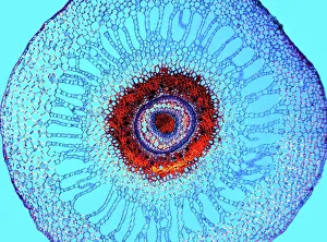
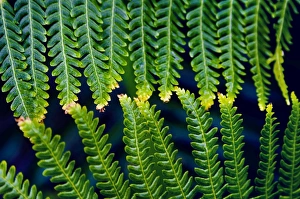
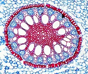
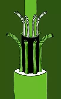
A vascular bundle, the intricate network of transport tissues in plants, is a fascinating subject to explore under the microscope


All Professionally Made to Order for Quick Shipping
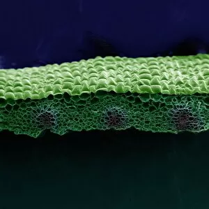
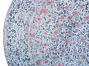
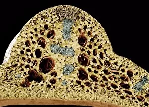
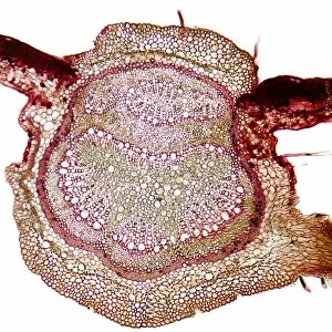
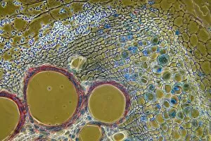
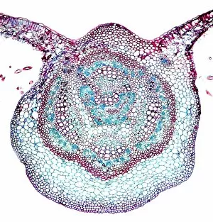
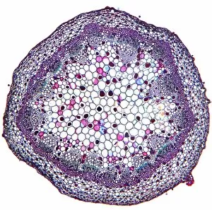
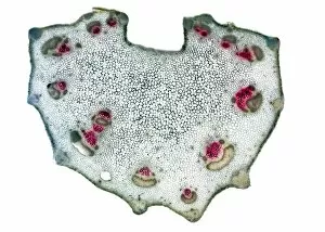
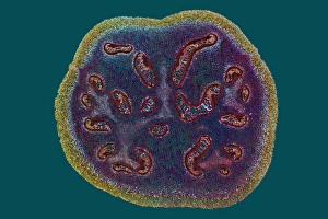
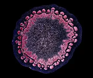
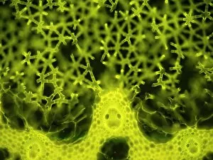
A vascular bundle, the intricate network of transport tissues in plants, is a fascinating subject to explore under the microscope. From the detailed SEM images of various plant parts like castor oil stem and maize root to the captivating light micrographs of water lily leaf and tomato leaf, these glimpses into nature's engineering marvels never cease to amaze. The water fern rhizome showcases its complex system for nutrient uptake while the pondweed stem reveals its efficient method for conducting water. In a mesmerizing display, fern leaves touch one another, highlighting their interconnectedness through vascular bundles. Delving deeper into this topic, an illustration depicts a typical plant vascular bundle with precision (C018 / 0915). Additionally, SEM images provide insight into dicotyledon vascular bundles (C016 / 9603 & C016 / 9602), showcasing their unique structures. As we examine these microscopic wonders like an iris root or dissected plant parts under bright lights (light micrograph), we gain a deeper appreciation for how plants efficiently distribute vital resources throughout their bodies using these remarkable vascular bundles.