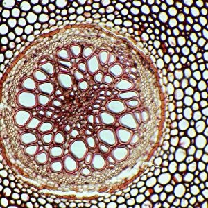
Fern rhizome, light micrograph

![]()
Wall Art and Photo Gifts from Science Photo Library
Fern rhizome, light micrograph
Fern rhizome. Polarised light micrograph of a cross-section through a rhizome from a male Dryopteris felix-mas fern. This is the central area of the rhizome, surrounded by bands of outer supporting fibres (red). In this central area are meristeles (oval areas) made up of vascular tissue. The meristele layers are: a single layer of endoderm cells (deep green); the cells of the pericycle and phloem (light green); and the large, open vessels of xylem (orange). Magnification: x100 when printed at 10 centimetres across
Science Photo Library features Science and Medical images including photos and illustrations
Media ID 6333259
© DR KEITH WHEELER/SCIENCE PHOTO LIBRARY
Cellular Cross Section Fern Nutrient Transport Phloem Polarised Light Micrograph Polarised Light Microscopy Polarized Rhizome Tissue Transverse Trio Vascular Bundle Vascular Bundles Water Transport Xylem Cells Light Micrograph Light Microscope Section Sectioned
MADE IN AUSTRALIA
Safe Shipping with 30 Day Money Back Guarantee
FREE PERSONALISATION*
We are proud to offer a range of customisation features including Personalised Captions, Color Filters and Picture Zoom Tools
SECURE PAYMENTS
We happily accept a wide range of payment options so you can pay for the things you need in the way that is most convenient for you
* Options may vary by product and licensing agreement. Zoomed Pictures can be adjusted in the Cart.