Fraud Blocker Skip to main content

Cancer Cells Collection

"Cancer Cells: Unveiling the Intricacies of a Silent Threat" Picture No

Background imageCancer Cells Collection: Picture No. 12479757

Picture No. 12479757
Zebrafish, Danio rerio, with human cancer. Zebrafish Date:

Background imageCancer Cells Collection: Picture No. 12479766

Picture No. 12479766
Zebrafish, Danio rerio. Veil fin variety above Date:

Background imageCancer Cells Collection: Picture No. 12479762

Picture No. 12479762
Newborn Zebrafish, Danio rerio. Zebrafish are Date:

Background imageCancer Cells Collection: Picture No. 12479764

Picture No. 12479764
Zebrafish, Danio rerio, fry on aquarium. Since Date:

Background imageCancer Cells Collection: Picture No. 12479761

Picture No. 12479761
Zebrafish, Danio rerio, used on cancer research. Date:

Background imageCancer Cells Collection: Picture No. 12479760

Picture No. 12479760
Zebrafish, Danio rerio. Stripe form (above) Casper Date:

Background imageCancer Cells Collection: Picture No. 12479765

Picture No. 12479765
Zebrafish, Danio rerio, fry on aquarium. Since Date:

Background imageCancer Cells Collection: Picture No. 12479763

Picture No. 12479763
Zebrafish, Danio rerio, fry on aquarium. Since Date:

Background imageCancer Cells Collection: Picture No. 12479756

Picture No. 12479756
Zebrafish, Danio rerio, with human cancer. Zebrafish Date:

Background imageCancer Cells Collection: Medical illustration depicting the different stages of colon cancer

Medical illustration depicting the different stages of colon cancer

Background imageCancer Cells Collection: Conceptual image of the Human Papilloma Virus

Conceptual image of the Human Papilloma Virus (HPV). HPV is a virus from the papillomavirus family that affects human skin and the moist membranes that line the body

Background imageCancer Cells Collection: Cancer cell with red blood cell flow

Cancer cell with red blood cell flow

Background imageCancer Cells Collection: Multiphoton fluorescence image of HeLa cells

Multiphoton fluorescence image of HeLa cells with cytoskeletal microtubules (magenta) and DNA (cyan)

Background imageCancer Cells Collection: Conceptual image of a tumor in human brain

Conceptual image of a tumor in human brain

Background imageCancer Cells Collection: Conceptual image of cancer virus

Conceptual image of cancer virus

Background imageCancer Cells Collection: Ewings sarcoma locations on the skeleton

Ewings sarcoma locations on the skeleton and detail of tumor on head of femur

Background imageCancer Cells Collection: Microscopic view of cancer virus

Microscopic view of cancer virus

Background imageCancer Cells Collection: Isolated cancer cell Macrophage

Isolated cancer cell Macrophage. Macrophages contribute to tumor growth and progression. Attracted to oxygen-starved (hypoxic) and necrotic tumor cells they promote chronic inflammation

Background imageCancer Cells Collection: Microscopic view of cancer cells

Microscopic view of cancer cells. Cancer occurs when a cells gene mutations make the cell unable to correct DNA damage

Background imageCancer Cells Collection: Conceptual image illustrating the process of bone metastasis

Conceptual image illustrating the process of bone metastasis

Background imageCancer Cells Collection: Microscopic view of tumor

Microscopic view of tumor

Background imageCancer Cells Collection: Microscopic view of pancreatic cancer cell

Microscopic view of pancreatic cancer cell

Background imageCancer Cells Collection: Microscipic view of pancreatic cancer cells

Microscipic view of pancreatic cancer cells

Background imageCancer Cells Collection: Scanning electron micrograph of an apoptotic HeLa cell

Scanning electron micrograph of an apoptotic HeLa cell. Zeiss Merlin HR-SEM


All Professionally Made to Order for Quick Shipping

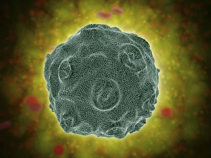
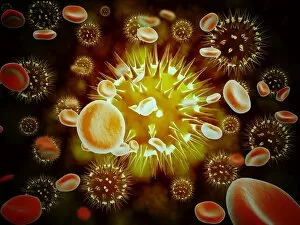
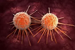
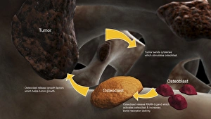
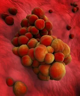
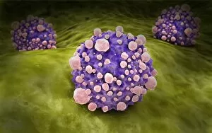
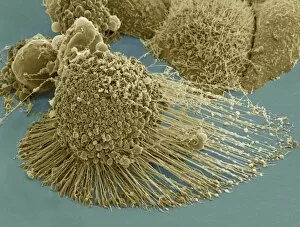
"Cancer Cells: Unveiling the Intricacies of a Silent Threat" Picture No. 12479757: A medical illustration depicting the different stages of colon cancer, showcasing the progression from benign polyps to malignant tumors. This visual representation highlights the importance of early detection and regular screenings. Picture No. 12479766: A microscopic view of a tumor reveals its intricate structure, composed of rapidly dividing cancer cells that invade surrounding tissues. Understanding these cellular abnormalities is crucial in developing targeted therapies for effective treatment. Picture No. 12479762: they can notorious for their ability to evade normal cell death mechanisms, leading to uncontrolled growth and proliferation. This image captures this relentless nature as cancer cells multiply uncontrollably, forming clusters within affected organs. Picture No. 12479764: The Human Papilloma Virus (HPV) has been linked to various types of cancers, including cervical and oral cancers. This conceptual image serves as a reminder that certain infections can contribute to the development of cancerous cells. Picture No. 12479761: Cancer research continues to unravel new insights into the molecular mechanisms driving tumor formation and progression. Scientists tirelessly investigate genetic mutations and signaling pathways within cancer cells with hopes of discovering breakthrough treatments. Picture No. 12479760: The battle against cancer involves an interdisciplinary approach where surgeons, oncologists, radiologists, researchers, and patients unite against this formidable adversary. Collaboration is key in finding innovative solutions towards improved patient outcomes. Picture No. 12479765: Despite advancements in treatment options such as chemotherapy or radiation therapy, some aggressive cancers remain challenging to eradicate completely due to their ability to adapt and develop resistance over time. Picture No. 12479763: Early detection plays a pivotal role in increasing survival rates for many types of cancers by allowing prompt intervention before metastasis occurs – highlighting the significance of routine screenings for individuals at risk or displaying concerning symptoms. Picture No. 12479756.