
Capsid Collection
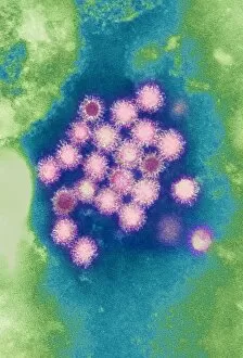
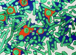
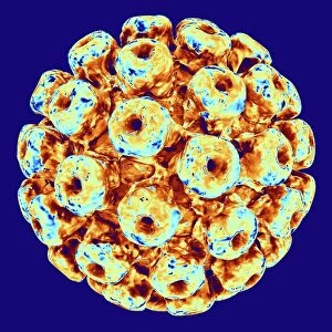
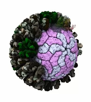
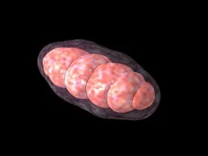
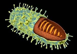
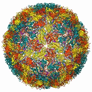
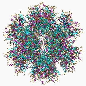
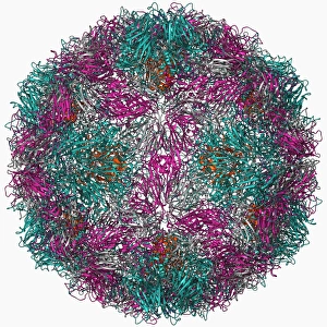
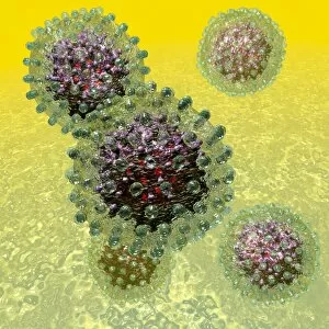
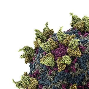
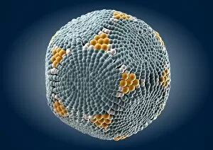
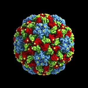
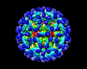
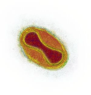
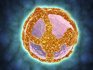
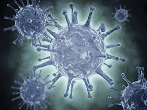
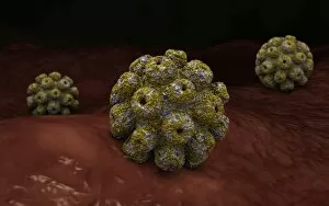
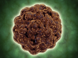
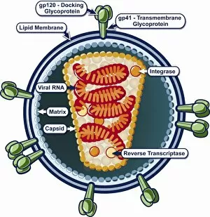
A capsid is a protective protein coat that surrounds the genetic material of viruses, such as Hepatitis B viruses and human respiratory syncytial virus


All Professionally Made to Order for Quick Shipping
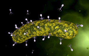
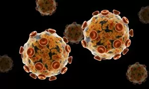
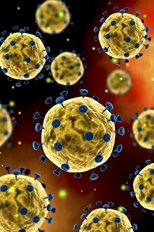
A capsid is a protective protein coat that surrounds the genetic material of viruses, such as Hepatitis B viruses and human respiratory syncytial virus. When viewed under a microscope, these tiny structures reveal intricate patterns and shapes that resemble works of art. For instance, the Adenovirus showcases its unique design in an artistic representation. The Hepatitis B viruses exhibit their characteristic capsids while replicating inside host cells. Similarly, Herpes virus particles can be observed undergoing replication through computer-generated artwork. Another example is the Polyoma BK virus, which displays its captivating structure in an artistic depiction. Computer-generated artwork also allows us to visualize the complex arrangement of Simian immunodeficiency virus (SIV) and Rotavirus particle structures. These images provide insight into their mechanisms of infection and replication. Bacteriophage phi29 is another fascinating example where a computer model helps us understand its unique shape and composition. This structure plays a crucial role in infecting bacteria. Through various forms of artwork, we gain appreciation for the intricacies within viral capsids. The beauty lies not only in their aesthetic appeal but also in unraveling their functions and understanding how they contribute to viral infections. Whether it's observing Hepatitis B viruses or admiring computer-generated representations of herpes virus particles, exploring different artworks showcasing diverse viral capsids offers valuable insights into virology research while simultaneously capturing our imagination with their stunning visuals.