Fraud Blocker Skip to main content

Cardiac Collection (page 2)

"Exploring the Intricacies of Cardiac Marvel

Background imageCardiac Collection: Climbing Cardiac Pass, Wasatch Mountains, Uinta Wasatch Cache National Forest, Utah

Climbing Cardiac Pass, Wasatch Mountains, Uinta Wasatch Cache National Forest, Utah
Climbing Cardiac Pass, Wasatch Mountains, Uinta-Wasatch-Cache National Forest, Utah

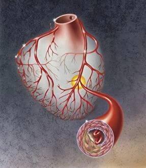
Background imageCardiac Collection: Heart with arteries showing cholesterol in one and plaque in the other

Heart with arteries showing cholesterol in one and plaque in the other
Heart with arteries showing cholesterol in one artery and atherosclerotic plaque in the other

Background imageCardiac Collection: Posterior view of human heart on photo of blood pressure cuff

Posterior view of human heart on photo of blood pressure cuff

Background imageCardiac Collection: Three dimensional view of human heart, right side

Three dimensional view of human heart, right side

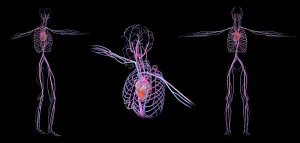
Background imageCardiac Collection: 3D rendering of human circulatory system

3D rendering of human circulatory system

Background imageCardiac Collection: Insertion of balloon into atherosclerotic artery

Insertion of balloon into atherosclerotic artery

Background imageCardiac Collection: Arteries on heart showing atherosclerotic plaque in an artery

Arteries on heart showing atherosclerotic plaque in an artery

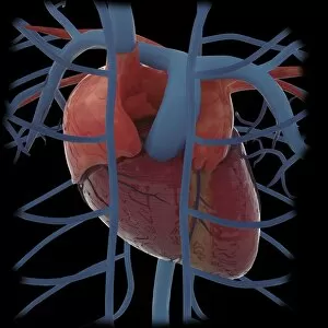
Background imageCardiac Collection: 3D rendering of human heart and thoracic veins

3D rendering of human heart and thoracic veins

Background imageCardiac Collection: Three dimensional view of human heart, back

Three dimensional view of human heart, back

Background imageCardiac Collection: Heart with pericadium pulled back exposing heart muscle

Heart with pericadium pulled back exposing heart muscle

Background imageCardiac Collection: Cross section of human heart

Cross section of human heart

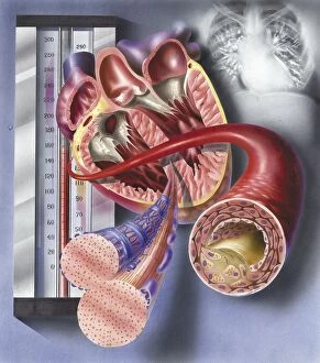
Background imageCardiac Collection: Interior view of heart with detail of muscle cells and atherosclerotic artery

Interior view of heart with detail of muscle cells and atherosclerotic artery. Chest x-ray in background

Background imageCardiac Collection: Detail of cardiac muscle with blood pressure meter

Detail of cardiac muscle with blood pressure meter

Background imageCardiac Collection: Human heart covered with pericardium

Human heart covered with pericardium

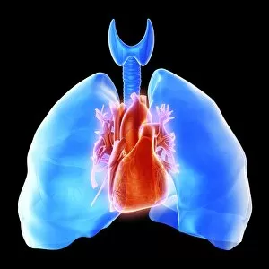
Background imageCardiac Collection: Pulmonary circulation of human heart and lung

Pulmonary circulation of human heart and lung

Background imageCardiac Collection: Heart valves showing pulmonary valve, mitral valve and tricuspid

Heart valves showing pulmonary valve, mitral valve and tricuspid

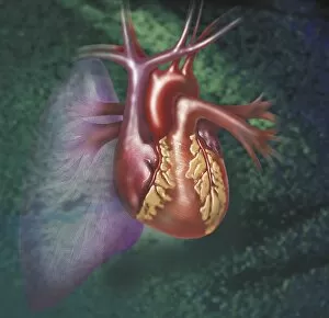
Background imageCardiac Collection: Anterior view of heart and lung on green background

Anterior view of heart and lung on green background

Background imageCardiac Collection: Comparison of normal heart versus heart with a patent foramen ovale

Comparison of normal heart versus heart with a patent foramen ovale

Background imageCardiac Collection: Conceptual image of human heart

Conceptual image of human heart

Background imageCardiac Collection: Internal view of the human heart

Internal view of the human heart

Background imageCardiac Collection: Interior of human heart showing atria and ventricles

Interior of human heart showing atria and ventricles

Background imageCardiac Collection: Enlarged left ventricle of the human heart

Enlarged left ventricle of the human heart

Background imageCardiac Collection: Three dimensional view of human heart, left side

Three dimensional view of human heart, left side

Background imageCardiac Collection: Medical illustration of human heart and rib cage

Medical illustration of human heart and rib cage, three dimensional view

Background imageCardiac Collection: Illustration of human heart

Illustration of human heart

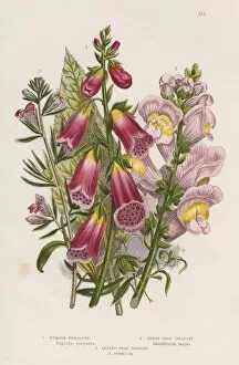
Background imageCardiac Collection: Foxglove (Digitalis purpurea), centre. The natural source of digitalin, a powerful

Foxglove (Digitalis purpurea), centre. The natural source of digitalin, a powerful cardiac stimulant, valuable in the treatment of heart disease

Background imageCardiac Collection: Model of a human heart

Model of a human heart

Background imageCardiac Collection: Healthy heart, artwork F005 / 0537

Healthy heart, artwork F005 / 0537
Healthy heart, computer artwork

Background imageCardiac Collection: ECG trace and head with brain, artwork

ECG trace and head with brain, artwork
ECG trace and brain. Computer artwork of an ECG (electrocardiogram) trace in front of a head and a human brain. An ECG uses electrodes to measure the electrical activity of the heart during

Background imageCardiac Collection: ECG trace and mri brain scan, artwork

ECG trace and mri brain scan, artwork
Brain anatomy. Coloured magnetic resonance imaging (MRI) scan of the human head from the side. The sagittal scan has halved the brain, revealing the internal anatomy

Background imageCardiac Collection: Healthy heart, artwork F005 / 0538

Healthy heart, artwork F005 / 0538
Healthy heart, computer artwork

Background imageCardiac Collection: Heartbeat trace, artwork C018 / 8585

Heartbeat trace, artwork C018 / 8585
Heartbeat trace. Artwork of the output trace from an electrocardiograph (ECG), a method used to monitor the beating of the heart

Background imageCardiac Collection: Heartbeat alert, artwork C018 / 8588

Heartbeat alert, artwork C018 / 8588
Heartbeat alert. Artwork of world alert with the output trace from an electrocardiograph (ECG), a method used to monitor the beating of the heart

Background imageCardiac Collection: Heartbeat trace, artwork C018 / 8586

Heartbeat trace, artwork C018 / 8586
Heartbeat trace. Artwork of the output trace from an electrocardiograph (ECG), a method used to monitor the beating of the heart

Background imageCardiac Collection: Heartbeat trace, artwork C018 / 8587

Heartbeat trace, artwork C018 / 8587
Heartbeat trace. Artwork of the output trace from an electrocardiograph (ECG), a method used to monitor the beating of the heart

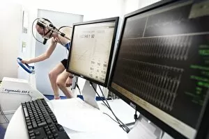
Background imageCardiac Collection: Physical fitness test C014 / 1103

Physical fitness test C014 / 1103
Physical fitness test. Athlete riding an exercise bike while his oxygen consumption is measured with a face mask. Electrodes on his chest monitor his heart rate

Background imageCardiac Collection: Physical fitness test C014 / 1108

Physical fitness test C014 / 1108
Physical fitness test. Athlete riding an exercise bike while his oxygen consumption is measured with a face mask. Electrodes on his chest monitor his heart rate (shown on screen)

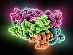
Background imageCardiac Collection: KCNQ ion channel protein structure

KCNQ ion channel protein structure. Molecular model showing the protein structure of an ion channel domain. Ion channels are membrane-spanning proteins that form a pathway for the movement of

Background imageCardiac Collection: Heart-lung anatomy, 1825 artwork

Heart-lung anatomy, 1825 artwork
Heart-lung anatomy. Axial section through the chest at the level of the fifth thoracic vertebra (T5). The front of the body is at top

Background imageCardiac Collection: Telemedicine, conceptual image C017 / 7589

Telemedicine, conceptual image C017 / 7589
Telemedicine, conceptual image

Background imageCardiac Collection: Physical fitness test C014 / 1105

Physical fitness test C014 / 1105
Physical fitness test. Athlete riding an exercise bike while his oxygen consumption is measured with a face mask. Electrodes on his chest monitor his heart rate

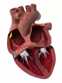
Background imageCardiac Collection: Heart chambers and sinus node, artwork C014 / 2030

Heart chambers and sinus node, artwork C014 / 2030
Heart chambers and sinus node (yellow). Cutaway artwork revealing the four internal heart chambers and the heart valves and tendons (white). The two upper chambers are the atria

Background imageCardiac Collection: Pulmonary hypertension, artwork

Pulmonary hypertension, artwork
Pulmonary hypertension, computer artwork. Pulmonary hypertension is a term to describe a disease in which the blood pressure in the lungs (pulmonary artery system) is higher than normal

Background imageCardiac Collection: 3-hydroxyacyl-CoA dehydrogenase F006 / 9602

3-hydroxyacyl-CoA dehydrogenase F006 / 9602
3-hydroxyacyl-CoA dehydrogenase, molecular model. This enzyme is found in human heart tissue, and catalyses a reaction that is part of the beta-oxidation pathway

Background imageCardiac Collection: Heart chamber anatomy, artwork C014 / 2029

Heart chamber anatomy, artwork C014 / 2029
Heart chamber anatomy. Cutaway artwork revealing the four internal heart chambers and the heart valves and tendons (white). The two upper chambers are the atria

Background imageCardiac Collection: Telemedicine, conceptual image C017 / 7588

Telemedicine, conceptual image C017 / 7588
Telemedicine, conceptual image

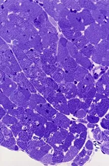
Background imageCardiac Collection: Cardiac muscle, light micrograph

Cardiac muscle, light micrograph
Cardiac muscle. Light micrograph of a section through muscle tissue from a heart, showing the cardiomyocyte cells. Heart muscle cells show central nuclei but the dominant feature is the abundance of

Background imageCardiac Collection: Atrial extrasystole, anatomical artwork

Atrial extrasystole, anatomical artwork
Atrial extrasystole. Anatomical computer artwork of a transverse section through a human heart during atrial contraction in extrasystole


All Professionally Made to Order for Quick Shipping

"Exploring the Intricacies of Cardiac Marvel: From Ancient Artwork to Modern Science" Delve into the captivating world wonders as we unravel the mysteries that lie within our beating hearts. The cardiovascular system, an intricate network responsible for sustaining life, has fascinated artists and scientists throughout history. Historical artwork depicting the human heart anatomy showcases early attempts to understand this vital organ's complexity. These masterpieces offer glimpses into how ancient civilizations perceived the seat of emotions and life itself. The irregular heartbeat, a condition that affects millions worldwide, is a subject both feared and studied. Captivating artwork portrays the chaotic dance of electrical impulses within our hearts, reminding us of its fragility and resilience. In mesmerizing depictions, artists capture not only the beauty but also the interconnectedness between our hearts and lungs—the symbiotic relationship that ensures oxygen-rich blood reaches every corner of our bodies. Witnessing red blood cells coursing through vessels towards their rendezvous with this mighty organ evokes awe at nature's ingenious design. Conceptual images portraying heart care remind us to nurture this precious entity with love and attention it deserves. Underneath a confocal light micrograph lies an enchanting sight—a glimpse into the intricate web of muscle fibers composing our heart muscle. This microscopic view reveals its strength in propelling life-sustaining blood throughout our bodies tirelessly. The human heart itself stands as a symbol revered across cultures—an emblematic representation transcending time and borders. Its significance extends beyond mere biology; it embodies love, compassion, courage—a vessel carrying profound emotions within each beat. Travel back centuries as we explore 16th-century anatomical drawings showcasing early endeavors to comprehend this enigmatic organ fully. These works serve as testaments to humanity's ceaseless quest for knowledge about ourselves. Artistic renditions capturing muscle contractions reveal dynamic movements hidden beneath layers upon layers—each contraction fueling existence itself—a symphony orchestrated by the heart's rhythmic beats.