Fraud Blocker Skip to main content

Cardiac Collection (page 7)

"Exploring the Intricacies of Cardiac Marvel

Background imageCardiac Collection: Artificial heart valve showing bacterial infection

Artificial heart valve showing bacterial infection
Bacterial endocarditis. Close-up of an infected artificial heart valve (Hancock type) showing bacterial endocarditis. (granulated tissue at centre)

Background imageCardiac Collection: Narrowed coronary artery

Narrowed coronary artery

Background imageCardiac Collection: Heart attack: artwork of heart angiogram and ECG

Heart attack: artwork of heart angiogram and ECG
Heart attack. Computer composite artwork depicting a heart attack (myocardial infarction), showing an angiogram of coronary arteries and an ECG heart trace

Background imageCardiac Collection: Angina pectoris

Angina pectoris. Illustration of a heart diseased with the cardiac condition angina pectoris. The heart is outlined in blue, with the aortic arch at top (red) and coronary arteries at lower frame

Background imageCardiac Collection: Man holds his chest due to angina or heart attack

Man holds his chest due to angina or heart attack
Angina or heart attack. Computer artwork of a man clutching his chest during angina pectoris or a heart attack. The image shows the heart muscle glowing yellow indicating pain

Background imageCardiac Collection: Heart disease: hand held up to irregular ECG trace

Heart disease: hand held up to irregular ECG trace
MODEL RELEASED. Heart disease. Computer artwork of a man holding his hand up to an ECG (electro- cardiogram) trace showing an irregular and falling heartbeat

Background imageCardiac Collection: Coloured SEM of a blood clot in coronary artery

Coloured SEM of a blood clot in coronary artery
Thrombus. Coloured Scanning Electron Micrograph (SEM) of a thrombus (blood clot) inside a coronary artery of the human heart

Background imageCardiac Collection: Angina or heart attack

Angina or heart attack
MODEL RELEASED. Angina or heart attack. Man clutching his chest in pain due to angina or a heart attack (myocardial infarction)

Background imageCardiac Collection: Heart disease: artwork of an irregular ECG trace

Heart disease: artwork of an irregular ECG trace

Background imageCardiac Collection: Colour SEM of atherosclerosis in coronary artery

Colour SEM of atherosclerosis in coronary artery
Atherosclerosis. Coloured Scanning Electron Micro- graph (SEM) of a longitudinal section through a human coronary artery of the heart showing athero- sclerosis

Background imageCardiac Collection: High blood pressure, artwork

High blood pressure, artwork
High blood pressure. Artwork of some of the effects of high blood pressure (hypertension). This can be caused by an excess of calcium ions (green, lower centre)

Background imageCardiac Collection: Artwork of heart in congestive heart failure

Artwork of heart in congestive heart failure
Heart failure. Cutaway illustration of a human heart showing congestive ventricular heart failure. Blood vessels connecting the heart to the rest of the body are at top

Background imageCardiac Collection: Artwork of human heart wrapped in drug information

Artwork of human heart wrapped in drug information
Heart disease & medication. Artwork of the human heart wrapped in text describing drugs prescribed to counteract heart disease or disorders of the heart

Background imageCardiac Collection: LM of section whole heart showing LV infarct

LM of section whole heart showing LV infarct
Infarcted left ventricle: light micrograph of a section through the ventricles of a whole human heart, showing an area of infarction (muscle necrosis or death)

Background imageCardiac Collection: Scintigram showing ischaemia post heart attack

Scintigram showing ischaemia post heart attack
False-colour scintigram (gamma camera scan) of a human heart showing ischaemia (reduced blood flow) following myocardial infarction

Background imageCardiac Collection: Artwork of silhouette with heart in brains place

Artwork of silhouette with heart in brains place
Heart neurosis. Artwork of a human profile in silhouette with a heart in the place of the brain, depicting heart neurosis. Heart (cardiac) neurosis is excessive fear of the condition of the heart

Background imageCardiac Collection: Illustration of blood pressure measurement

Illustration of blood pressure measurement. Blood pressure is a measurement of the pressure that blood exerts against the walls of the main arteries and is quoted in millimetres of mercury (mmHg)

Background imageCardiac Collection: Atrial fibrillation, artwork

Atrial fibrillation, artwork
Atrial fibrillation. Artwork of a section through a human heart during atrial fibrillation, a rapid, irregular heartbeat. Two electrocardiogram traces (ECGs) are seen at centre left and upper right

Background imageCardiac Collection: Abstract comp. graphics of fading human pulse

Abstract comp. graphics of fading human pulse
Heart failure: abstract computer graphics image based on a fading electrocardiogram (ECG) trace, a record of the electrical activity within the heart as it beats

Background imageCardiac Collection: Artwork showing atherosclerosis of coronary artery

Artwork showing atherosclerosis of coronary artery
Coronary artery disease. Illustration of a human heart showing narrowing of the coronary artery due to atherosclerosis - build up of fatty deposits (atheroma, yellow)

Background imageCardiac Collection: Artwork of heart & ECG trace in heart failure

Artwork of heart & ECG trace in heart failure
Heart failure. Cutaway illustration of a human heart and an ECG (electrocardiogram) trace in left ventricular heart failure

Background imageCardiac Collection: Coloured X-ray of heart showing dextrocardia

Coloured X-ray of heart showing dextrocardia
Heart dextrocardia. Coloured X-ray of the chest of a patient showing dextrocardia, a rare condition where the heart is situated in the right hand side of the chest (at left on image)

Background imageCardiac Collection: Light micrograph of cardiac muscle & fatty disease

Light micrograph of cardiac muscle & fatty disease

Background imageCardiac Collection: False-col chest x-ray showing hypertension

False-col chest x-ray showing hypertension
False-colour chest x-ray in a case of hypertension, abnormally high blood pressure, showing an enlarged heart at bottom right obscurring the lower lobe of the left lung

Background imageCardiac Collection: Heart disease: hypertension, atheroma, lung oedema

Heart disease: hypertension, atheroma, lung oedema

Background imageCardiac Collection: Artwork of heart attack due to atherosclerosis

Artwork of heart attack due to atherosclerosis
Heart attack. Artwork of a human heart during a heart attack (myocardial infarction). Here, an embolus (red, in the sectioned artery at lower right)

Background imageCardiac Collection: Illustration of a ventricular hole in the heart

Illustration of a ventricular hole in the heart. This hole in the septum between the two ventricles is a heart abnormality developed before birth (congenital)

Background imageCardiac Collection: Conceptual artwork of heart with heart disease

Conceptual artwork of heart with heart disease
Heart disease. Conceptual artwork of a section through a heart to depict it " running down" due to heart disease

Background imageCardiac Collection: Coloured chest X-ray of ventricular cardiomyopathy

Coloured chest X-ray of ventricular cardiomyopathy

Background imageCardiac Collection: Heart disease: art of heart & beta blocker symbol

Heart disease: art of heart & beta blocker symbol
Heart disease and beta blocker drugs. Artwork of a heart and a symbol representing the use of beta blocker drugs. Beta blockers are used to treat angina, to lower blood pressure

Background imageCardiac Collection: Heart, chest X-ray

Heart, chest X-ray

Background imageCardiac Collection: Contour map of the heart

Contour map of the heart, computer artwork. The contours on this map describe the surface shape of the heart, shown here from the front

Background imageCardiac Collection: Pericardium, TEM

Pericardium, TEM
Pericardium. Coloured transmission electron micrograph (TEM) of a section through the serous pericardium. The pericardium is a double-walled sac surrounding the heart

Background imageCardiac Collection: Heart

Heart, wire-frame computer artwork. The heart is a muscular pump that circulates blood around the body, providing all tissues with oxygen and nutrients

Background imageCardiac Collection: Heart

Heart with lines behind it, computer artwork. The heart is a hollow muscle that pumps blood around the body. Deoxygenated blood from the body is carried into the right side of the heart

Background imageCardiac Collection: Heart of gold

Heart of gold, computer artwork. The heart is a hollow sac (lower frame) of muscular tissue that pumps blood around the body. Small surface coronary blood vessels supply the heart muscle with blood

Background imageCardiac Collection: Heart

Heart and glowing light, computer artwork. The heart (red) is a hollow sac of muscular tissue that pumps blood around the body

Background imageCardiac Collection: Heart valve tendons, SEM

Heart valve tendons, SEM
Heart valve tendons. Coloured scanning electron micrograph (SEM) of the chordae tendineae, tendons which attach the atrioventricular heart valves to their muscles

Background imageCardiac Collection: Heart of glass

Heart of glass, computer artwork. The heart is a hollow muscle that pumps blood around the body. The vena cava vein (upper left) carries deoxygenated blood into the heart from the body

Background imageCardiac Collection: Heart

Heart. Computer artwork of a heart with a barrier around it. This could represent the protection of the heart against heart disease or further heart failure by drugs such as ACE inhibitors

Background imageCardiac Collection: Heart blood vessels, SEM

Heart blood vessels, SEM
Heart blood vessels. Coloured scanning electron micrograph (SEM) of a freeze-fractured section through cardiac muscle, exposing the blood vessels of the heart

Background imageCardiac Collection: Protected heart, conceptual artwork

Protected heart, conceptual artwork
Protected heart, conceptual computer artwork. The metal cage around the heart could represent protection of the heart against heart disease or further heart failure, by drugs such as ACE inhibitors

Background imageCardiac Collection: Heart and red blood cells, artwork

Heart and red blood cells, artwork
Heart and red blood cells, computer artwork. The heart is a hollow muscle that pumps blood around the body. Deoxygenated blood from the body is carried into the right side of the heart (left)

Background imageCardiac Collection: Electrical conduction in the heart, artwork

Electrical conduction in the heart, artwork. Each heartbeat is initiated by an electrical impulse from the sinoatrial (SA) node (green oval) in the right atrium (upper heart chamber)

Background imageCardiac Collection: Dissected heart, light micrograph

Dissected heart, light micrograph
Dissected heart. Light micrograph of a section through a whole heart, showing the chambers and some blood. The heart is a hollow muscular organ that pumps blood around the body

Background imageCardiac Collection: Heart and lightbeams

Heart and lightbeams
Heart with beams of light behind it, computer artwork. The heart is a hollow muscle that pumps blood around the body. Deoxygenated blood from the body is carried into the right side of the heart

Background imageCardiac Collection: ECG and red blood cells

ECG and red blood cells. Computer artwork of an electrocardiogram (ECG) trace and red blood cells (erythrocytes). Red blood cells are biconcave, giving them a large surface area for gas exchange

Background imageCardiac Collection: Heart and red blood cells

Heart and red blood cells (erythrocytes), computer artwork. The coronary blood vessels are seen surrounding the heart. The heart is a muscular pump that moves blood around the body


All Professionally Made to Order for Quick Shipping

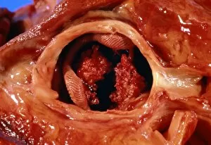
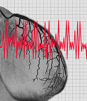
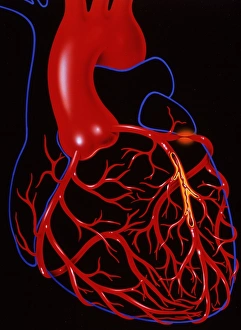
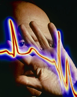
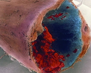
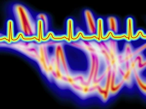
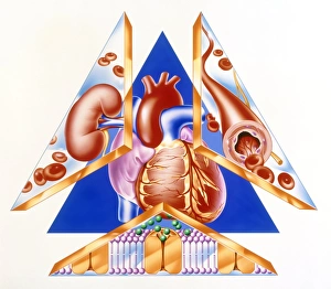
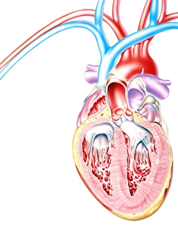
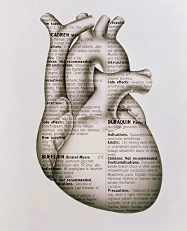
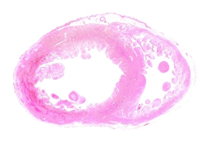
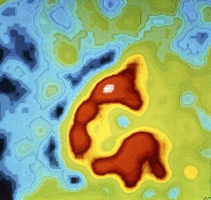
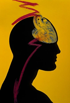
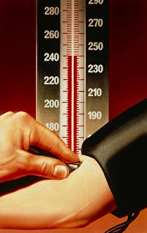
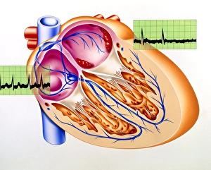
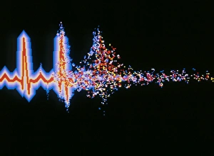
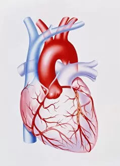
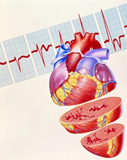
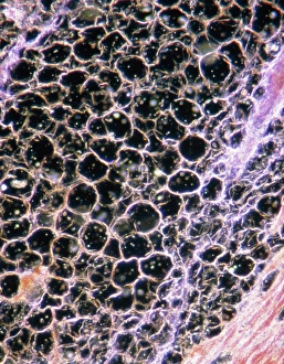
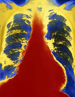
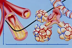
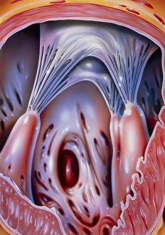
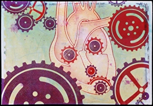
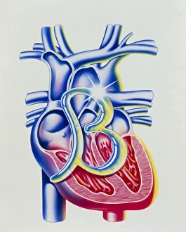
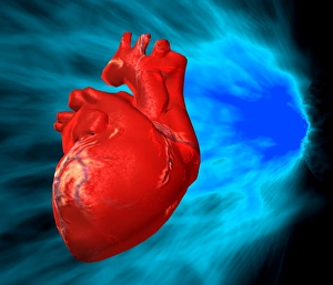
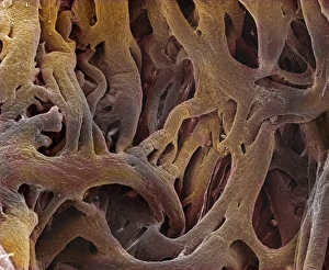
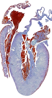
"Exploring the Intricacies of Cardiac Marvel: From Ancient Artwork to Modern Science" Delve into the captivating world wonders as we unravel the mysteries that lie within our beating hearts. The cardiovascular system, an intricate network responsible for sustaining life, has fascinated artists and scientists throughout history. Historical artwork depicting the human heart anatomy showcases early attempts to understand this vital organ's complexity. These masterpieces offer glimpses into how ancient civilizations perceived the seat of emotions and life itself. The irregular heartbeat, a condition that affects millions worldwide, is a subject both feared and studied. Captivating artwork portrays the chaotic dance of electrical impulses within our hearts, reminding us of its fragility and resilience. In mesmerizing depictions, artists capture not only the beauty but also the interconnectedness between our hearts and lungs—the symbiotic relationship that ensures oxygen-rich blood reaches every corner of our bodies. Witnessing red blood cells coursing through vessels towards their rendezvous with this mighty organ evokes awe at nature's ingenious design. Conceptual images portraying heart care remind us to nurture this precious entity with love and attention it deserves. Underneath a confocal light micrograph lies an enchanting sight—a glimpse into the intricate web of muscle fibers composing our heart muscle. This microscopic view reveals its strength in propelling life-sustaining blood throughout our bodies tirelessly. The human heart itself stands as a symbol revered across cultures—an emblematic representation transcending time and borders. Its significance extends beyond mere biology; it embodies love, compassion, courage—a vessel carrying profound emotions within each beat. Travel back centuries as we explore 16th-century anatomical drawings showcasing early endeavors to comprehend this enigmatic organ fully. These works serve as testaments to humanity's ceaseless quest for knowledge about ourselves. Artistic renditions capturing muscle contractions reveal dynamic movements hidden beneath layers upon layers—each contraction fueling existence itself—a symphony orchestrated by the heart's rhythmic beats.