
Colonic Collection
"Exploring the Depths: Unveiling the Importance Health" Discovering Harrogate for Health and Pleasure, a haven for wellness enthusiasts seeking internal rejuvenation


All Professionally Made to Order for Quick Shipping
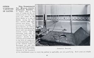
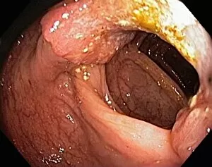
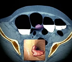
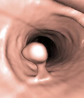
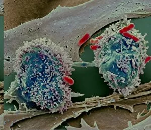
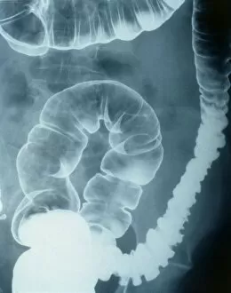
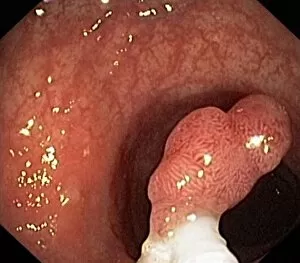
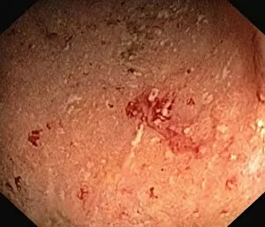
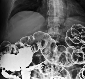
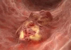
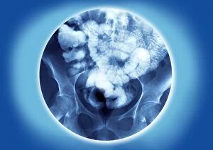
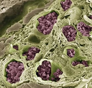
"Exploring the Depths: Unveiling the Importance Health" Discovering Harrogate for Health and Pleasure, a haven for wellness enthusiasts seeking internal rejuvenation. Delve into the world treatments, where ancient practices meet modern technology. Beneath captivating black and white photos lies the transformative power of an internal douche - a refreshing cleanse that revitalizes your body from within. Witness how intestinal hernias can be understood through mesmerizing artwork (C016 / 7525), shedding light on this common condition. Uncover the hidden truths behind constipation with X-ray revelations, showcasing its impact on our digestive system. Barium contrast CT scans expose colon cancer's presence, reminding us to prioritize regular screenings as early detection saves lives. Embark on a visual journey through 3D colonoscopy images, revealing intricate details of both colon cancer and polyps. These cutting-edge technologies provide invaluable insights into prevention and treatment options. Witness once again barium contrast CT scans capturing colon cancer's relentless nature while realizing that vigilance is key in combating this disease. Marvel at microscopic wonders with a light micrograph (C016 / 0522) unveiling the intricacies of our large intestine. In this exploration health, we are reminded that maintaining optimal well-being goes beyond surface-level care. Embrace these hints as gentle reminders to prioritize your inner health – because true vitality starts from within.