Fraud Blocker Skip to main content

Compound Collection (page 17)

A fascinating world of compounds unfolds in this captivating collection of images

Background imageCompound Collection: Auranofin drug molecule

Auranofin drug molecule. Computer artwork showing the structure of a molecule of the drug auranofin. Auranofin, marketed under the name Ridaura

Background imageCompound Collection: Melatonin, light micrograph

Melatonin, light micrograph
Melatonin. Light micrograph in polarised light of crystals of the hormone melatonin. Melatonin is secreted by the pineal gland in the brain that controls the bodys biological rhythm

Background imageCompound Collection: Vitamin B1 molecule

Vitamin B1 molecule. Computer model showing the structure of a molecule of vitamin B1 (thiamine). Vitamin B1 is an essential nutrient that humans are unable to produce

Background imageCompound Collection: Testosterone hormone molecule

Testosterone hormone molecule. Computer model showing the structure of a molecule of the male sex hormone testosterone. Testosterone is the main human androgen

Background imageCompound Collection: Progesterone hormone molecule

Progesterone hormone molecule. Computer model showing the structure of a molecule of the hormone progesterone. Progesterone is produced in the ovaries of women and the testes of men

Background imageCompound Collection: Glyphosate weed killer molecule

Glyphosate weed killer molecule. Computer model showing the molecular structure of a molecule of the herbicide glyphosate. Glyphosate is a widely used herbicide

Background imageCompound Collection: Ibuprofen molecule

Ibuprofen molecule. Computer artwork showing the structure of a molecule of the painkilling (analgesic) drug ibuprofen. Ibuprofen is a non-steroidal anti-inflammatory drug (NSAID)

Background imageCompound Collection: Graphene sheet

Graphene sheet. Computer artwork showing the structure of a graphene sheet. Graphene is a single layer of graphite. It is composed of hexagonally arranged carbon atoms (spheres)

Background imageCompound Collection: Caffeine molecule

Caffeine molecule. Computer artwork showing the structure of a molecule of the alkaloid stimulant and legal drug caffeine. Caffeine is found in drinks such as tea, coffee, and fizzy drinks

Background imageCompound Collection: Asbestos fibres, light micrograph

Asbestos fibres, light micrograph
Light micrograph with Normarski Differential Contrast (DIC) illumination of asbestos fibres. Asbestos has been used in the building industry for many years as a result of its heat resistance

Background imageCompound Collection: Serotonin molecule

Serotonin molecule. Computer model showing the structure of a molecule of the neurotransmitter (nerve signalling chemical) serotonin (5-hydroxytryptamine)

Background imageCompound Collection: Aspirin molecule

Aspirin molecule. Computer artwork showing the structure of a molecule of aspirin (acetylsalicylic acid). Atoms are represented as spheres and are colour-coded: carbon (black), hydrogen (white)

Background imageCompound Collection: Aspirin in action

Aspirin in action. Computer artwork showing how aspirin has its effect. Aspirin (acetylsalicylic acid) is converted to salicylic acid and acetic acid in the body

Background imageCompound Collection: Testosterone hormone, light micrograph C015 / 6787

Testosterone hormone, light micrograph C015 / 6787
Testosterone hormone. Polarised light micrograph of crystals of the male sex hormone testosterone. Testosterone is the main human androgen hormone

Background imageCompound Collection: Coagulation factor complex molecule C014 / 0626

Coagulation factor complex molecule C014 / 0626
Coagulation factor complex molecule. Molecular model showing a blood clotting factor VIIa-tissue factor complex molecule (blue and white ribbons) with a tissue factor pathway inhibitor (TFPI)

Background imageCompound Collection: Compound leaves, 19th century C015 / 6084

Compound leaves, 19th century C015 / 6084
Compound leaves, 19th-century artwork. Compound leaves are those where the leaf is divided into separate leaflets, as opposed to the single blade or lobes of simple leaves (see C015/6083)

Background imageCompound Collection: Coagulation factor complex molecule C014 / 0585

Coagulation factor complex molecule C014 / 0585
Coagulation factor complex molecule. Molecular model showing a blood clotting factor VIIa-tissue factor complex molecule (blue and white) with a tissue factor pathway inhibitor (TFPI)

Background imageCompound Collection: Coagulation factor complex molecule C014 / 0584

Coagulation factor complex molecule C014 / 0584
Coagulation factor complex molecule. Molecular model showing a blood clotting factor VIIa-tissue factor complex molecule (blue and white ribbons) with a tissue factor pathway inhibitor (TFPI)

Background imageCompound Collection: Westinghouse electric generator, 1897

Westinghouse electric generator, 1897
Westinghouse electric generator. 19th-century artwork of a compound-type generator (right) directly coupled to a dynamo (left) by a flexible sleeve

Background imageCompound Collection: Preparing a medicinal iodine solution C018 / 2316

Preparing a medicinal iodine solution C018 / 2316
Pharmaceutics. A technician pouring a prepared iodine solution into small bottles for medicinal use. Iodine is used in antiseptic compounds such as povidone-iodine

Background imageCompound Collection: Insulin crystals, light micrograph C017 / 8246

Insulin crystals, light micrograph C017 / 8246
Insulin. Polarised light micrograph (PLM) of crystals of the hormone insulin. The insulin molecule is made up of two chains of amino acids (A & B chains)

Background imageCompound Collection: Oestrogen receptor, molecular model

Oestrogen receptor, molecular model
Oestrogen receptor. Molecular model of an oestrogen receptor, bound to a glucocorticoid receptor-interacting protein. Oestrogen receptors are cytoplasmic proteins that bind oestrogens

Background imageCompound Collection: Myoglobin molecule C015 / 5164

Myoglobin molecule C015 / 5164
Myoglobin molecule. Computer model showing the structure of a myoglobin molecule. Myoglobin is a protein found in muscle tissue

Background imageCompound Collection: Transcription factor and DNA molecule C015 / 5344

Transcription factor and DNA molecule C015 / 5344
Transcription factor and DNA molecule. Molecular model of glucocorticoid receptor (GR) transcription factor protein (blue and purple) complexed with a molecule of DNA (deoxyribonucleic acid)

Background imageCompound Collection: Transcription factor and DNA molecule C015 / 5343

Transcription factor and DNA molecule C015 / 5343
Transcription factor and DNA molecule. Molecular model of glucocorticoid receptor (GR) transcription factor protein (yellow and green) complexed with a molecule of DNA (deoxyribonucleic acid)

Background imageCompound Collection: Potassium iodide crystals, micrograph C016 / 3119

Potassium iodide crystals, micrograph C016 / 3119
Potassium iodide crystals, light micrograph. Potassium iodide (KI) is produced in large quantities industrially, and is used in a variety of products, such as disinfectants

Background imageCompound Collection: DNA and tumour suppressor complex C014 / 0014

DNA and tumour suppressor complex C014 / 0014
DNA and tumour suppressor complex. Molecular model showing a molecule of the p53 tumour suppressor protein (green, centre) bound to a damaged DNA (deoxyribonucleic acid) strand

Background imageCompound Collection: DNA and methyltransferase complex C014 / 0011

DNA and methyltransferase complex C014 / 0011
DNA and methyltransferase complex. Molecular model showing a molecule of methyltransferase bound to a DNA (deoxyribonucleic acid) strand (red and yellow)

Background imageCompound Collection: Neuropeptide Y neurotransmitter molecule C014 / 0013

Neuropeptide Y neurotransmitter molecule C014 / 0013
Neuropeptide Y neurotransmitter molecule. Molecular model showing the structure of the neurotransmitter neuropeptide Y (NPY)

Background imageCompound Collection: HRas enzyme molecule C014 / 0010

HRas enzyme molecule C014 / 0010
HRas enzyme molecule. Molecular model showing the structure of a molecule of the enzyme GTPase HRas, also known as transforming protein p21

Background imageCompound Collection: NADH dehydrogenase molecule, artwork C014 / 0009

NADH dehydrogenase molecule, artwork C014 / 0009
NADH dehydrogenase molecule. Computer artwork showing the structure of a molecule of NADH dehydrogenase (NADH:ubiquinone reductase or Complex I)

Background imageCompound Collection: Water molecule, artwork C014 / 0007

Water molecule, artwork C014 / 0007
Water molecule. Computer artwork showing the structure of a molecule of water (chemical formula H2O), consisting of two atoms of hydrogen (grey) bonded to one atom of oxygen (red)

Background imageCompound Collection: Benzene molecule, artwork

Benzene molecule, artwork
Benzene molecule, molecular model. Benzene is an aromatic organic compound that consists of a ring of six carbon atoms, each with an attached hydrogen atom

Background imageCompound Collection: Topiramate antiepileptic drug molecule C013 / 9956

Topiramate antiepileptic drug molecule C013 / 9956
Topiramate. Molecular model of the antiepileptic drug Topiramate (brand name Topamax), an anticonvulsant drug that decreases abnormal excitement in the brain

Background imageCompound Collection: Phentermine drug molecule C013 / 9952

Phentermine drug molecule C013 / 9952
Phentermine. Molecular model of the appetite suppressant phentermine. This drug is used to help obese patients lose weight by decreasing their appetite

Background imageCompound Collection: Human Apolipoprotein A-1 molecule

Human Apolipoprotein A-1 molecule. Molecular model showing the structure of a high-density lipoprotein (HDL) known as an apolipoprotein

Background imageCompound Collection: HIV reverse transcription enzyme C013 / 9613

HIV reverse transcription enzyme C013 / 9613
HIV reverse transcription enzyme. Molecular model of the reverse transcriptase enzyme found in HIV (the human immunodeficiency virus)

Background imageCompound Collection: 4-Methylimidazole molecule C013 / 9438

4-Methylimidazole molecule C013 / 9438
4-Methylimidazole molecule. Computer model showing the structure of a molecule of the heterocyclic organic chemical compound 4-Methylimidazole (4-MEI)

Background imageCompound Collection: 4-Methylimidazole molecule C013 / 9436

4-Methylimidazole molecule C013 / 9436
4-Methylimidazole molecule. Computer model showing the structure of a molecule of the heterocyclic organic chemical compound 4-Methylimidazole (4-MEI)

Background imageCompound Collection: Ribonuclease with RNA DNA hybrid

Ribonuclease with RNA DNA hybrid
Ribonuclease with RNA/DNA hybrid. Molecular model of Ribonuclease H (RNAse H, yellow and green) complexed with an RNA (ribonucleic acid, purple) and DNA (deoxyribonucleic acid, pink) hybrid


All Professionally Made to Order for Quick Shipping

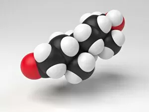
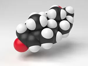
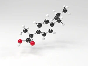
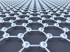
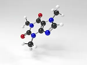
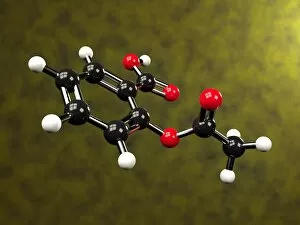
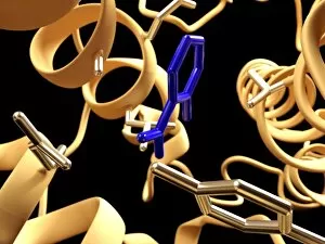
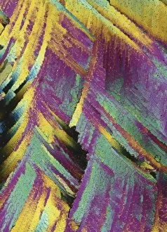
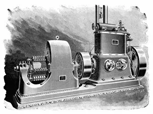
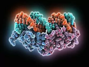
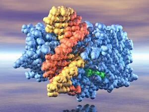
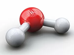
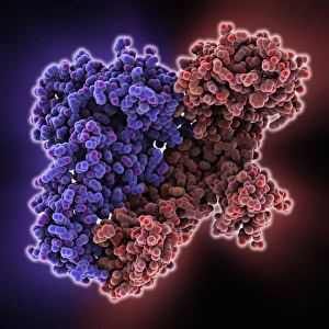
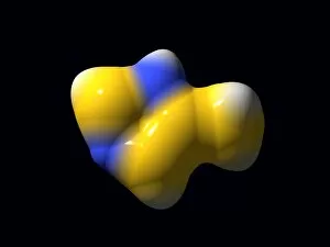
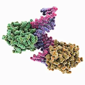
A fascinating world of compounds unfolds in this captivating collection of images. From the ancient artistry of knapped flint tools to the intricate beauty of copper and magnesium sulphate crystals, each compound tells its own unique story. In a mesmerizing light micrograph, caffeine crystals shimmer with delicate intricacy, while the process of threshing wheat showcases the compound's vital role in agriculture. The stunning EDTA crystals reveal their exquisite structure under microscopic examination, offering a glimpse into their molecular composition. Oxytocin hormone crystals take center stage in another breathtaking light micrograph, showcasing nature's ability to create intricate patterns. Meanwhile, an ant captured through scanning electron microscopy reminds us that even tiny creatures are composed of complex compounds. Artwork depicting secondary structures of proteins invites contemplation on the building blocks that make life possible. The perovskite crystal structure captivates with its geometric precision and potential for technological advancements. The Simulium damnosum, also known as the Simulian blackfly, is showcased as a remarkable example of nature's interconnectedness and reliance on compounds for survival. Light micrographs unveil oxytocin crystals once again, highlighting their ethereal beauty. Finally, a honey bee captured through scanning electron microscopy serves as a reminder that compounds shape not only our surroundings but also play an essential role in sustaining life itself. This captivating journey through various compounds offers glimpses into both natural wonders and human ingenuity. It underscores how these fundamental elements shape our world in ways both seen and unseen – from ancient tools to modern technology – reminding us that everything around us is ultimately comprised of compounds waiting to be discovered and understood.