Fraud Blocker Skip to main content

Compounds Collection (page 6)

"Exploring the Fascinating World of Compounds: From Copper and Magnesium Sulphate to Graphene" Delving into the intricate beauty of compounds

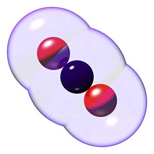
Background imageCompounds Collection: Carbon dioxide molecule C016 / 8493

Carbon dioxide molecule C016 / 8493
Carbon dioxide molecule. Computer artwork showing the structure of a molecule of carbon dioxide. Carbon dioxide is a colourless gas that occurs naturally in the atmosphere

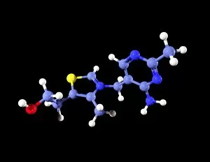
Background imageCompounds Collection: Vitamin B1 molecule C016 / 8278

Vitamin B1 molecule C016 / 8278
Vitamin B1 molecule. Computer model showing the structure of a molecule of vitamin B1 (thiamine). Atoms are represented as colour-coded spheres: carbon (light blue), hydrogen (white)

Background imageCompounds Collection: Vitamin B1 molecule C016 / 8277

Vitamin B1 molecule C016 / 8277
Vitamin B1 molecule. Computer model showing the structure of a molecule of vitamin B1 (thiamine). Vitamin B1 is an essential nutrient that humans are unable to produce

Background imageCompounds Collection: Vitamin B1 molecule C016 / 8276

Vitamin B1 molecule C016 / 8276
Vitamin B1 molecule. Computer model showing the structure of a molecule of vitamin B1 (thiamine). Vitamin B1 is an essential nutrient that humans are unable to produce

Background imageCompounds Collection: Vitamin B1 molecule C016 / 8275

Vitamin B1 molecule C016 / 8275
Vitamin B1 molecule. Computer model showing the structure of a molecule of vitamin B1 (thiamine). Vitamin B1 is an essential nutrient that humans are unable to produce

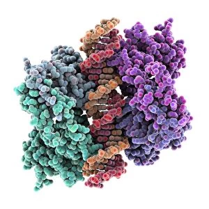
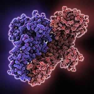
Background imageCompounds Collection: Tumour suppressor protein molecular model C016 / 2065

Tumour suppressor protein molecular model C016 / 2065
Tumour suppressor protein. Molecular model of the tumour suppressor protein p53 (left and right) bound to a molecule of DNA (deoxyribonucleic acid, down centre) at the p53 response element

Background imageCompounds Collection: Melatonin, light micrograph

Melatonin, light micrograph
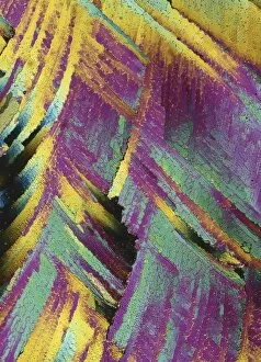
Melatonin. Light micrograph in polarised light of crystals of the hormone melatonin. Melatonin is secreted by the pineal gland in the brain that controls the bodys biological rhythm

Background imageCompounds Collection: Vitamin B1 molecule

Vitamin B1 molecule. Computer model showing the structure of a molecule of vitamin B1 (thiamine). Vitamin B1 is an essential nutrient that humans are unable to produce

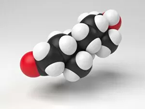
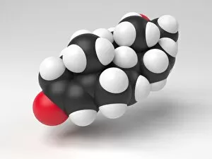
Background imageCompounds Collection: Testosterone hormone molecule

Testosterone hormone molecule. Computer model showing the structure of a molecule of the male sex hormone testosterone. Testosterone is the main human androgen

Background imageCompounds Collection: Progesterone hormone molecule

Progesterone hormone molecule. Computer model showing the structure of a molecule of the hormone progesterone. Progesterone is produced in the ovaries of women and the testes of men
Background imageCompounds Collection: Glyphosate weed killer molecule

Glyphosate weed killer molecule. Computer model showing the molecular structure of a molecule of the herbicide glyphosate. Glyphosate is a widely used herbicide

Background imageCompounds Collection: Ibuprofen molecule

Ibuprofen molecule. Computer artwork showing the structure of a molecule of the painkilling (analgesic) drug ibuprofen. Ibuprofen is a non-steroidal anti-inflammatory drug (NSAID)

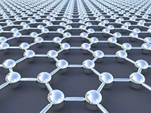
Background imageCompounds Collection: Graphene sheet

Graphene sheet. Computer artwork showing the structure of a graphene sheet. Graphene is a single layer of graphite. It is composed of hexagonally arranged carbon atoms (spheres)

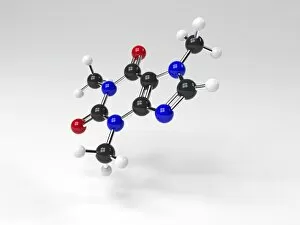
Background imageCompounds Collection: Caffeine molecule

Caffeine molecule. Computer artwork showing the structure of a molecule of the alkaloid stimulant and legal drug caffeine. Caffeine is found in drinks such as tea, coffee, and fizzy drinks

Background imageCompounds Collection: Asbestos fibres, light micrograph

Asbestos fibres, light micrograph
Light micrograph with Normarski Differential Contrast (DIC) illumination of asbestos fibres. Asbestos has been used in the building industry for many years as a result of its heat resistance

Background imageCompounds Collection: Serotonin molecule

Serotonin molecule. Computer model showing the structure of a molecule of the neurotransmitter (nerve signalling chemical) serotonin (5-hydroxytryptamine)

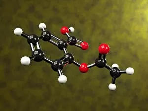
Background imageCompounds Collection: Aspirin molecule

Aspirin molecule. Computer artwork showing the structure of a molecule of aspirin (acetylsalicylic acid). Atoms are represented as spheres and are colour-coded: carbon (black), hydrogen (white)

Background imageCompounds Collection: Aspirin in action

Aspirin in action. Computer artwork showing how aspirin has its effect. Aspirin (acetylsalicylic acid) is converted to salicylic acid and acetic acid in the body

Background imageCompounds Collection: Testosterone hormone, light micrograph C015 / 6787

Testosterone hormone, light micrograph C015 / 6787
Testosterone hormone. Polarised light micrograph of crystals of the male sex hormone testosterone. Testosterone is the main human androgen hormone

Background imageCompounds Collection: Insulin crystals, light micrograph C017 / 8246

Insulin crystals, light micrograph C017 / 8246
Insulin. Polarised light micrograph (PLM) of crystals of the hormone insulin. The insulin molecule is made up of two chains of amino acids (A & B chains)

Background imageCompounds Collection: Oestrogen receptor, molecular model

Oestrogen receptor, molecular model
Oestrogen receptor. Molecular model of an oestrogen receptor, bound to a glucocorticoid receptor-interacting protein. Oestrogen receptors are cytoplasmic proteins that bind oestrogens

Background imageCompounds Collection: Myoglobin molecule C015 / 5164

Myoglobin molecule C015 / 5164
Myoglobin molecule. Computer model showing the structure of a myoglobin molecule. Myoglobin is a protein found in muscle tissue

Background imageCompounds Collection: Chemical Symbols

Chemical Symbols
Pictographic symbols used at the beginning of the 19th century to represent chemical elements and compounds. They are similar to those of the ancient alchemists

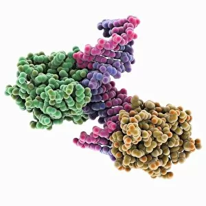
Background imageCompounds Collection: Transcription factor and DNA molecule C015 / 5344

Transcription factor and DNA molecule C015 / 5344
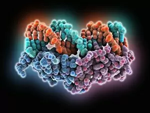
Transcription factor and DNA molecule. Molecular model of glucocorticoid receptor (GR) transcription factor protein (blue and purple) complexed with a molecule of DNA (deoxyribonucleic acid)

Background imageCompounds Collection: Transcription factor and DNA molecule C015 / 5343

Transcription factor and DNA molecule C015 / 5343
Transcription factor and DNA molecule. Molecular model of glucocorticoid receptor (GR) transcription factor protein (yellow and green) complexed with a molecule of DNA (deoxyribonucleic acid)

Background imageCompounds Collection: DNA and tumour suppressor complex C014 / 0014

DNA and tumour suppressor complex C014 / 0014
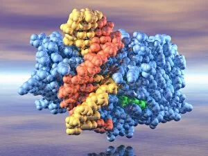
DNA and tumour suppressor complex. Molecular model showing a molecule of the p53 tumour suppressor protein (green, centre) bound to a damaged DNA (deoxyribonucleic acid) strand

Background imageCompounds Collection: DNA and methyltransferase complex C014 / 0011

DNA and methyltransferase complex C014 / 0011
DNA and methyltransferase complex. Molecular model showing a molecule of methyltransferase bound to a DNA (deoxyribonucleic acid) strand (red and yellow)

Background imageCompounds Collection: Neuropeptide Y neurotransmitter molecule C014 / 0013

Neuropeptide Y neurotransmitter molecule C014 / 0013
Neuropeptide Y neurotransmitter molecule. Molecular model showing the structure of the neurotransmitter neuropeptide Y (NPY)

Background imageCompounds Collection: HRas enzyme molecule C014 / 0010

HRas enzyme molecule C014 / 0010
HRas enzyme molecule. Molecular model showing the structure of a molecule of the enzyme GTPase HRas, also known as transforming protein p21

Background imageCompounds Collection: NADH dehydrogenase molecule, artwork C014 / 0009

NADH dehydrogenase molecule, artwork C014 / 0009
NADH dehydrogenase molecule. Computer artwork showing the structure of a molecule of NADH dehydrogenase (NADH:ubiquinone reductase or Complex I)

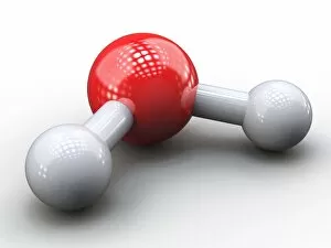
Background imageCompounds Collection: Water molecule, artwork C014 / 0007

Water molecule, artwork C014 / 0007
Water molecule. Computer artwork showing the structure of a molecule of water (chemical formula H2O), consisting of two atoms of hydrogen (grey) bonded to one atom of oxygen (red)

Background imageCompounds Collection: Benzene molecule, artwork

Benzene molecule, artwork
Benzene molecule, molecular model. Benzene is an aromatic organic compound that consists of a ring of six carbon atoms, each with an attached hydrogen atom

Background imageCompounds Collection: Topiramate antiepileptic drug molecule C013 / 9956

Topiramate antiepileptic drug molecule C013 / 9956
Topiramate. Molecular model of the antiepileptic drug Topiramate (brand name Topamax), an anticonvulsant drug that decreases abnormal excitement in the brain

Background imageCompounds Collection: Human Apolipoprotein A-1 molecule

Human Apolipoprotein A-1 molecule. Molecular model showing the structure of a high-density lipoprotein (HDL) known as an apolipoprotein

Background imageCompounds Collection: HIV reverse transcription enzyme C013 / 9613

HIV reverse transcription enzyme C013 / 9613
HIV reverse transcription enzyme. Molecular model of the reverse transcriptase enzyme found in HIV (the human immunodeficiency virus)

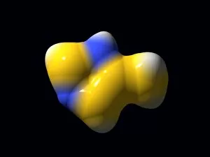
Background imageCompounds Collection: 4-Methylimidazole molecule C013 / 9438

4-Methylimidazole molecule C013 / 9438
4-Methylimidazole molecule. Computer model showing the structure of a molecule of the heterocyclic organic chemical compound 4-Methylimidazole (4-MEI)

Background imageCompounds Collection: 4-Methylimidazole molecule C013 / 9436

4-Methylimidazole molecule C013 / 9436
4-Methylimidazole molecule. Computer model showing the structure of a molecule of the heterocyclic organic chemical compound 4-Methylimidazole (4-MEI)

Background imageCompounds Collection: Ribonuclease with RNA DNA hybrid

Ribonuclease with RNA DNA hybrid
Ribonuclease with RNA/DNA hybrid. Molecular model of Ribonuclease H (RNAse H, yellow and green) complexed with an RNA (ribonucleic acid, purple) and DNA (deoxyribonucleic acid, pink) hybrid


All Professionally Made to Order for Quick Shipping

"Exploring the Fascinating World of Compounds: From Copper and Magnesium Sulphate to Graphene" Delving into the intricate beauty of compounds, we witness the mesmerizing sight of copper and magnesium sulphate crystals under a light microscope (LM). A closer look at caffeine crystals through a light micrograph reveals their captivating structure, resembling tiny jewels that fuel our mornings. Oxytocin hormone crystals, captured using polarized light microscopy (PLM C016 / 7196), unveil the remarkable complexity behind this molecule responsible for human bonding. Through an artistic representation, we unravel the secondary structure of proteins – nature's building blocks that orchestrate countless biological processes within us. The perovskite crystal structure captivates scientists with its potential applications in renewable energy technologies, promising a brighter future for sustainable power generation. Another glimpse into oxytocin's world showcases its crystalline form under a light microscope, reminding us of its vital role in nurturing social connections and maternal instincts. Zooming in on caffeine's molecular composition unveils its drug-like qualities that stimulate our nervous system and keep us awake during long nights or early mornings. Peering into the microscopic realm reveals bacterial ribosomes - miniature protein factories essential for life itself - showcasing nature's incredible machinery at work. Cortisol crystals come to life as they are illuminated by a beam of light under a microscope, offering insight into this stress hormone's unique properties within our bodies. Exploring vitamin B12 through its molecular model highlights how this crucial nutrient supports various bodily functions while displaying an elegant arrangement of atoms and bonds. Once again, copper sulphate crystals enchant us with their vibrant colors when observed using a light microscope (LM), reminding us of their diverse industrial uses and chemical significance. Stepping into the realm of materials science brings forth graphene.