Fraud Blocker Skip to main content

Compounds Collection (page 8)

"Exploring the Fascinating World of Compounds: From Copper and Magnesium Sulphate to Graphene" Delving into the intricate beauty of compounds

Background imageCompounds Collection: Human growth hormone molecule C013 / 9006

Human growth hormone molecule C013 / 9006
Human growth hormone. Molecular model of human growth hormone (hGH, yellow) bound to the extracellular domain of the human growth hormone binding protein (hGHBP)

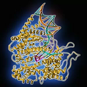
Background imageCompounds Collection: RNA polymerase molecule C013 / 9005

RNA polymerase molecule C013 / 9005
RNA polymerase. Molecular model of RNA polymerase (yellow) transcribing a strand of mRNA (messenger ribonucleic acid, pink) from a DNA (deoxyribonucleic acid) template (orange and turquoise)

Background imageCompounds Collection: HIV reverse transcription enzyme C013 / 8998

HIV reverse transcription enzyme C013 / 8998
HIV reverse transcription enzyme. Molecular model of the reverse transcriptase enzyme (orange and blue) found in HIV (the human immunodeficiency virus)

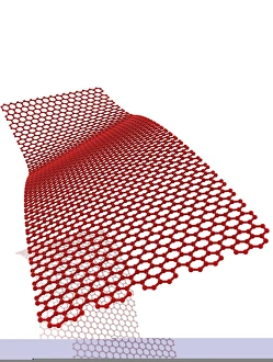
Background imageCompounds Collection: Graphene conductivity, conceptual image

Graphene conductivity, conceptual image. Computer artwork of a graphene sheet with electricity (yellow) passing through it. Graphene is a single layer of graphite

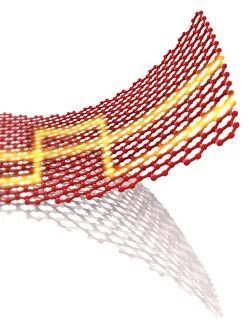
Background imageCompounds Collection: Graphene conductivity, conceptual image C013 / 8897

Graphene conductivity, conceptual image C013 / 8897
Graphene conductivity, conceptual image. Computer artwork of a graphene sheet with electricity (yellow) passing through it. Graphene is a single layer of graphite

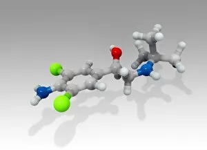
Background imageCompounds Collection: Clenbuterol bronchodilator drug molecule C013 / 8893

Clenbuterol bronchodilator drug molecule C013 / 8893
Clenbuterol drug molecule. This drug is a decongestant and bronchodilator used to treat breathing disorders. It acts on beta-2 adrenergic receptors in the smooth muscle of the airways

Background imageCompounds Collection: Erythropoietin molecule C013 / 8891

Erythropoietin molecule C013 / 8891
Erythropoietin molecule (EPO), molecular model. EPO is a glycoprotein that is produced by the kidneys in response to low blood oxygen levels

Background imageCompounds Collection: Birch pollen allergen molecule C013 / 8889

Birch pollen allergen molecule C013 / 8889
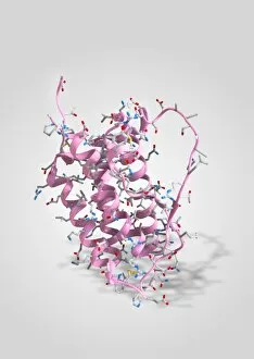
Birch pollen allergen molecule. Computer model showing the secondary structure of a Bet v 1L molecule. This molecule is responsible for allergic reactions to pollen from birch (Betula sp.) trees

Background imageCompounds Collection: Green fluorescent protein molecule C013 / 8885

Green fluorescent protein molecule C013 / 8885
Green fluorescent protein molecule. Computer model showing the secondary structure of a molecule of green fluorescent protein (GFP). GFP is found in the Pacific jellyfish Aequorea victoria

Background imageCompounds Collection: Bovine adrenodoxin molecules C013 / 8876

Bovine adrenodoxin molecules C013 / 8876
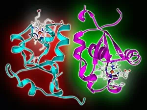
Bovine adrenodoxin molecules. Computer models showing the secondary structure of two molecules of adrenodoxin (adrenal ferredoxin) from a cow

Background imageCompounds Collection: Cytochrome b5 molecules C013 / 8874

Cytochrome b5 molecules C013 / 8874
Cytochrome b5 molecules. Computer artwork showing the secondary structure of two molecules of cytochrome B5. This protein plays a crucial role in metabolism in animals (including humans), fungi

Background imageCompounds Collection: Tumour suppressor protein molecular model C013 / 7914

Tumour suppressor protein molecular model C013 / 7914
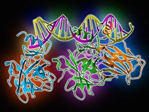
Tumour suppressor protein. Molecular model of the tumour suppressor protein p53 (blue, green and orange) bound to a molecule of DNA (deoxyribonucleic acid, yellow and pink)

Background imageCompounds Collection: DNA polymerase molecule C013 / 7909

DNA polymerase molecule C013 / 7909
DNA polymerase. Molecular model of a molecule of DNA polymerase (blue) replicating a strand of DNA (deoxyribonucleic acid, pink and turquoise). The secondary structure of the DNA polymerase is shown

Background imageCompounds Collection: Ascorbic acid crystals (SEM) C013 / 7831

Ascorbic acid crystals (SEM) C013 / 7831
Ascorbic acid crystals or vitamin C, showing the monoclinic crystal structure of the pure form (characterised by 3 unequal axes, one pair of which are not at right angles)

Background imageCompounds Collection: Abcorbic acid crystals (SEM) C013 / 7830

Abcorbic acid crystals (SEM) C013 / 7830
Ascorbic acid crystals or vitamin C, showing the monoclinic crystal structure of the pure form (characterised by 3 unequal axes, one pair of which are not at right angles)

Background imageCompounds Collection: Gypsum crystals SEM

Gypsum crystals SEM
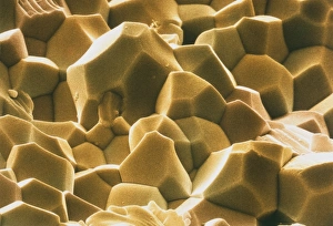
Gypsum crystals. Coloured scanning electron micrograph (SEM) of crystals of calcium sulphate dihydrate, or gypsum. Gypsum is used as plaster of Paris

Background imageCompounds Collection: Rose quartz crystals, SEM

Rose quartz crystals, SEM
Rose quartz crystals, coloured scanning electron micrograph (SEM). Quartz is a form of silica (silicon dioxide) and the most abundant mineral in the Earths crust

Background imageCompounds Collection: Coloured SEM of beryllium oxide crystals

Coloured SEM of beryllium oxide crystals
Beryllium oxide crystals. Coloured scanning electron micrograph (SEM) of the fractured surface of beryllium oxide (BeO) ceramic

Background imageCompounds Collection: High density foam, Zotefoam

High density foam, Zotefoam
Zotefoam. Coloured scanning electron micrograph of Zotefoam LD45, a high density synthetic foam. Zotefoam is characterized by the relatively uniform size of the cells (irregular shapes)

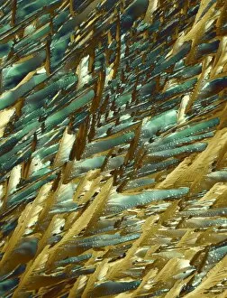
Background imageCompounds Collection: TEMPUR memory foam, SEM

TEMPUR memory foam, SEM
TEMPUR memory foam, coloured scanning electron micrograph (SEM). This viscoelastic memory foam, made from polyurethane, was originally developed in the 1970s at NASA Ames Research Center, California

Background imageCompounds Collection: Reflective belt material

Reflective belt material
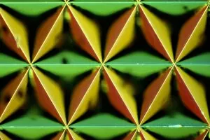
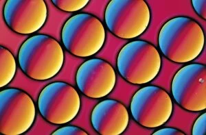
Light micrograph in interference contrast illumination of a reflective material used in safety clothing. The material is retro-reflective

Background imageCompounds Collection: Resin used in solid phase peptide synthesis A850 / 0042

Resin used in solid phase peptide synthesis A850 / 0042
Light micrograph of p-Methylbenzhydrylamine, an inert resin used in solid phase peptide synthesis; an amide group is attached to the resin allowing further amino acids to be built up in a chain

Background imageCompounds Collection: SEM of silver crystals

SEM of silver crystals
Silver crystals. Coloured scanning electron micrograph (SEM) of dendritic (branching, tree- like) crystals of silver (symbol Ag, atomic number 47)

Background imageCompounds Collection: Crystals of silver on copper wire

Crystals of silver on copper wire
Light micrograph in incident illumination of crystalline silver deposited on a copper wire. Copper wire is suspended in a solution of silver nitrate

Background imageCompounds Collection: LM of melted arsenic disulphide

LM of melted arsenic disulphide
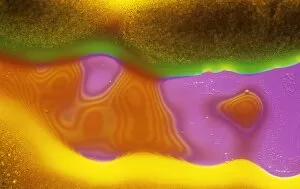
Light micrograph of molten arsenic disulphide. Interference contrast. Magnification: x200 at 35mm size

Background imageCompounds Collection: Calcium sulphate crystals, SEM

Calcium sulphate crystals, SEM
Calcium sulphate crystals. Coloured scanning electron micrograph (SEM) of crystals of calcium sulphate (CaSO4), or gypsum. Calcium sulphate is used as plaster of Paris

Background imageCompounds Collection: Light micrograph of picric acid crystals

Light micrograph of picric acid crystals
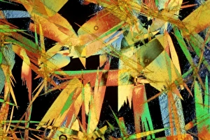
Light micrograph in interference contrast illumination of picric acid crystals. Picric acid is an explosive, and is commonly used in home-made bombs. Magnification x200 at 35mm size

Background imageCompounds Collection: Kettle fur limescale

Kettle fur limescale
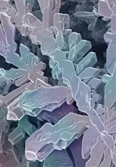
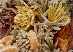
Kettle fur". Coloured scanning electron micrograph (SEM) of fur from a domestic kettle, showing the crystals that form in a hard water area

Background imageCompounds Collection: LM of silver nitrate crystals

LM of silver nitrate crystals
Polarised light micrograph of silver nitrate crystals. Silver nitrate is used in photography and as a test for chloride, bromide and iodide ions. Magnification: x100 at 35mm size

Background imageCompounds Collection: Radiochemical Centre

Radiochemical Centre
Process boxes for the production of tritium compounds at the Radiochemical Centre, Amersham, Buckinghamshire, England. Date: December 1964

Background imageCompounds Collection: Water quality scientist from Thames Water, taking samples to be tested for traces of metaldehyde

Water quality scientist from Thames Water, taking samples to be tested for traces of metaldehyde, carbetamide and propyzamide, River Cherwell, Cropredy Bridge, Oxfordshire, England, march

Background imageCompounds Collection: Red blood cells and molecules, artwork

Red blood cells and molecules, artwork
Red blood cells and drug molecules, computer artwork. Red blood cells (erythrocytes) are responsible for supplying tissues with oxygen and are the most abundant type of cell in the blood

Background imageCompounds Collection: Wolcott Gibbs, US chemist

Wolcott Gibbs, US chemist
Wolcott Gibbs (1822-1908), US chemist. Gibbs graduated from Columbia University, USA in 1841 and qualified as a medical doctor in 1845

Background imageCompounds Collection: Carbon nanotube and buckyball, artwork

Carbon nanotube and buckyball, artwork
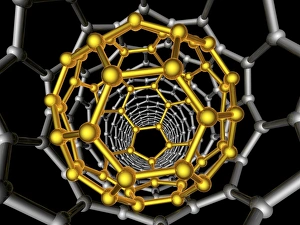
Carbon nanotube and buckyball. Computer artwork showing the hexagonal carbon structures of a buckyball (yellow) inside a carbon nanotube, or buckytube (grey)

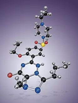
Background imageCompounds Collection: Levitra drug molecule

Levitra drug molecule
Levitra. Molecular model of the drug vardenafil, marketed under the brand Levitra. Atoms are represented as spheres and are colour-coded: carbon (black), hydrogen (silver), nitrogen (blue)

Background imageCompounds Collection: Cialis drug molecule

Cialis drug molecule
Cialis. Molecular model of the drug tadalafil, marketed under the brand Cialis. Atoms are represented as spheres and are colour-coded: carbon (black), hydrogen (grey), nitrogen (blue), oxygen (red)

Background imageCompounds Collection: Fluoxetine drug molecule

Fluoxetine drug molecule
Fluoxetine. Molecular model of the drug fluoxetine, which is marketed under the brand Prozac. Atoms are represented as spheres and are colour-coded: carbon (black), hydrogen (silver)

Background imageCompounds Collection: Viagra drug molecule

Viagra drug molecule
Viagra. Molecular model of the drug sildenafil citrate, marketed under the brands Viagra and Revatio. Atoms are represented as spheres and are colour-coded: carbon (black), hydrogen (silver)

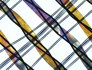
Background imageCompounds Collection: Synthetic fibre, light micrograph

Synthetic fibre, light micrograph
Light micrograph of a synthetic fibre, taken with polarised light. Magnification: x100

Background imageCompounds Collection: Glycine crystals, light micrograph

Glycine crystals, light micrograph
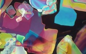
Glycine crystals. Polarised light micrograph of crystals of glycine, the simplest of the amino acids. Glycine consists of a central carbon atom, to which are attached two hydrogen atoms

Background imageCompounds Collection: Adrenaline, polarized light micrograph

Adrenaline, polarized light micrograph
Adrenaline. Polarized light micrograph of crystals of adrenaline (epinephrine). This is a hormone produced in the adrenal glands above the kidneys


All Professionally Made to Order for Quick Shipping

"Exploring the Fascinating World of Compounds: From Copper and Magnesium Sulphate to Graphene" Delving into the intricate beauty of compounds, we witness the mesmerizing sight of copper and magnesium sulphate crystals under a light microscope (LM). A closer look at caffeine crystals through a light micrograph reveals their captivating structure, resembling tiny jewels that fuel our mornings. Oxytocin hormone crystals, captured using polarized light microscopy (PLM C016 / 7196), unveil the remarkable complexity behind this molecule responsible for human bonding. Through an artistic representation, we unravel the secondary structure of proteins – nature's building blocks that orchestrate countless biological processes within us. The perovskite crystal structure captivates scientists with its potential applications in renewable energy technologies, promising a brighter future for sustainable power generation. Another glimpse into oxytocin's world showcases its crystalline form under a light microscope, reminding us of its vital role in nurturing social connections and maternal instincts. Zooming in on caffeine's molecular composition unveils its drug-like qualities that stimulate our nervous system and keep us awake during long nights or early mornings. Peering into the microscopic realm reveals bacterial ribosomes - miniature protein factories essential for life itself - showcasing nature's incredible machinery at work. Cortisol crystals come to life as they are illuminated by a beam of light under a microscope, offering insight into this stress hormone's unique properties within our bodies. Exploring vitamin B12 through its molecular model highlights how this crucial nutrient supports various bodily functions while displaying an elegant arrangement of atoms and bonds. Once again, copper sulphate crystals enchant us with their vibrant colors when observed using a light microscope (LM), reminding us of their diverse industrial uses and chemical significance. Stepping into the realm of materials science brings forth graphene.