Fraud Blocker Skip to main content

Cortex Collection (page 3)

The cortex, a fascinating part of the brain, is responsible for numerous functions that are essential to our daily lives

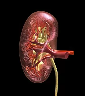
Background imageCortex Collection: Kidney anatomy, artwork C016 / 8663

Kidney anatomy, artwork C016 / 8663
Kidney anatomy, artwork. The kidneys filter waste from the blood and excrete it as urine. The kidney consists of an outer cortex and an inner medulla

Background imageCortex Collection: Kidney anatomy, artwork C016 / 8661

Kidney anatomy, artwork C016 / 8661
Kidney anatomy, artwork. The kidneys filter waste from the blood and excrete it as urine. The kidney consists of an outer cortex and an inner medulla

Background imageCortex Collection: Kidney anatomy, artwork C016 / 8662

Kidney anatomy, artwork C016 / 8662
Kidney anatomy, artwork. The kidneys filter waste from the blood and excrete it as urine. The kidney consists of an outer cortex and an inner medulla

Background imageCortex Collection: Kidney anatomy, artwork C016 / 8660

Kidney anatomy, artwork C016 / 8660
Kidney anatomy, artwork. The kidneys filter waste from the blood and excrete it as urine. The kidney consists of an outer cortex and an inner medulla

Background imageCortex Collection: Paprosky femur defect, type IIIA lateral

Paprosky femur defect, type IIIA lateral
Paprosky femur defect. Cutaway artwork of bone degradation in a type IV medial-lateral femur cortex defect (Paprosky classification system)

Background imageCortex Collection: Paprosky femur defect, type IIIA med-lat

Paprosky femur defect, type IIIA med-lat
Paprosky femur defect. Cutaway artwork of bone degradation in a type IV lateral femur cortex defect (Paprosky classification system)

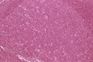
Background imageCortex Collection: Lymph node hyperplasia, light micrograph C015 / 6224

Lymph node hyperplasia, light micrograph C015 / 6224
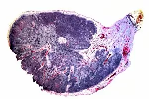
Lymph node hyperplasia. Light micrograph of a section through a lymph node showing follicular hyperplasia. Hyperplasia is the abnormal proliferation of cells

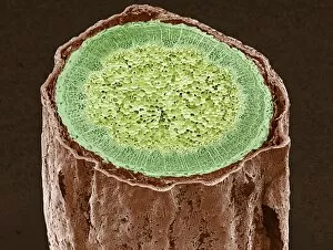
Background imageCortex Collection: Magnolia stem, SEM C015 / 5128

Magnolia stem, SEM C015 / 5128
Magnolia stem. Coloured environmental scanning electron micrograph (ESEM) of a transverse section through the stem of a Magnolia sp. plant

Background imageCortex Collection: Magnolia stem, SEM C015 / 5124

Magnolia stem, SEM C015 / 5124
Magnolia stem. Coloured environmental scanning electron micrograph (ESEM) of a transverse section through the stem of a Magnolia sp. plant

Background imageCortex Collection: Lymph node, artwork C013 / 4632

Lymph node, artwork C013 / 4632
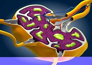
Lymph node. Computer artwork of a section through a human lymph node. Lymph nodes are kidney-shaped structures located at intervals along lymph vessels (tubes)

Background imageCortex Collection: Lymph node, artwork C013 / 4631

Lymph node, artwork C013 / 4631
Lymph node. Computer artwork of a section through a human lymph node. Lymph nodes are kidney-shaped structures located at intervals along lymph vessels (tubes)

Background imageCortex Collection: Artwork of the structure of a human hair

Artwork of the structure of a human hair

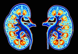
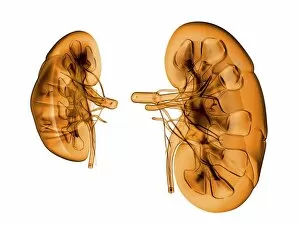
Background imageCortex Collection: Human kidneys

Human kidneys. Computer artwork of two sectioned human kidneys. Humans possess two kidneys, located at the back of the abdomen, responsible for excreting nitrogenous waste from the blood

Background imageCortex Collection: Kidney

Kidney

Background imageCortex Collection: LM of section through cerebral cortex and medulla

LM of section through cerebral cortex and medulla
Cerebrum. Light micrograph of a section through the cerebrum of a human brain showing the cortex and medulla. The cerebrum is formed of two lobes (left and right)

Background imageCortex Collection: Cross-section of cortex & medulla of cerebellum

Cross-section of cortex & medulla of cerebellum

Background imageCortex Collection: Brain nerve cells

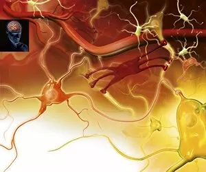
Brain nerve cells
Nerve cells in the brain. Artwork showing the different types of nerve cells in the grey matter of the brain. Neurons (yellow, for example at lower right) relay nerve signals around the brain

Background imageCortex Collection: Human brain, occipital lobe

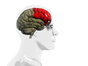
Human brain, occipital lobe
Brain, temporal lobe. Computer artwork of a human brain in profile within a glass-effect male head, showing the location of the temporal lobe (red)

Background imageCortex Collection: Contour map of the brain

Contour map of the brain, computer artwork. The contours on this map describe the uneven surface the cortex of the brain, shown here from the left side

Background imageCortex Collection: Human brain, temporal lobe

Human brain, temporal lobe
Brain, brainstem. Computer artwork of a human brain in profile within a glass-effect female head, showing the location of the brainstem and central brain structures (red)

Background imageCortex Collection: SEM of lymphocytes in cortex of thymus

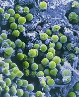
SEM of lymphocytes in cortex of thymus
False-colour scanning electron micrograph of the cortex of a thymus. The spheres are T-lymphocytes, white blood cells vital to the cell mediated resp- onse of the immune system

Background imageCortex Collection: Computer graphic of section through human kidney

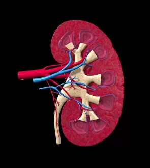
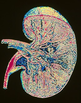
Computer graphic of section through human kidney
Kidney. Computer graphic of a section through a whole human kidney. At upper left is the renal artery and vein which supply blood to, and drain blood from the kidney

Background imageCortex Collection: Immunofluorescent LM of rat brain cells and axons

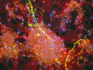
Immunofluorescent LM of rat brain cells and axons
Cortical brain cells and axons. Immunofluorescent Light Micrograph of rat cortical brain cells and axons growing in culture

Background imageCortex Collection: Granule nerve cell, SEM

Granule nerve cell, SEM
Granule nerve cell. Coloured scanning electron micrograph (SEM) of a granule nerve cell (yellow) from the cerebellum of the brain

Background imageCortex Collection: Brain

Brain. Computer artwork of a healthy human brain seen from above. The front of the brain is at top right. This is the cerebrum, the largest part of the brain

Background imageCortex Collection: Cerebellum, light micrograph

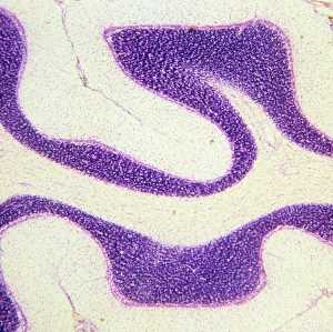
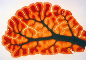
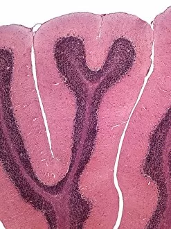
Cerebellum, light micrograph
Cerebellum. Light micrograph of a section through the cerebellum of the brain. The cerebellum comprises three main layers

Background imageCortex Collection: Human brain, conceptual artwork

Human brain, conceptual artwork
Human brain, conceptual computer artwork. The front of the brain is at right

Background imageCortex Collection: Kidneys, artwork

Kidneys, artwork
Kidneys, computer artwork

Background imageCortex Collection: Cerebellum structure, light micrograph

Cerebellum structure, light micrograph
Cerebellum structure. Coloured light micrograph of a section through the highly-folded cerebellum of the brain. The cerebellum comprises three main layers

Background imageCortex Collection: Brain scans

Brain scans. Sagittal (sideways) scan through the head, revealing the brain structure. The front of the head is at left. The scan shows the convoluted folds of the outer region

Background imageCortex Collection: Iris stem, light micrograph

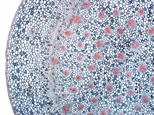
Iris stem, light micrograph. A thin outer epidermis (far left) surrounds a thick layer of cortex. In the centre of the stem is the pith, which contains numerous vascular bundles (red)

Background imageCortex Collection: Common rush stem, light micrograph

Common rush stem, light micrograph
Common rush stem. Light micrograph of a section through the stem of a common rush (Juncus conglomeratus) plant, showing stellate cells

Background imageCortex Collection: Dendrobium orchid root, light micrograph

Dendrobium orchid root, light micrograph
Dendrobium orchid root. Light micrograph of a section through an aerial root from a Dendrobium sp. orchid. The outer tissue (velamen radicum, grey) is composed of hexagonal cells

Background imageCortex Collection: Thymus gland tissue, light micrograph

Thymus gland tissue, light micrograph
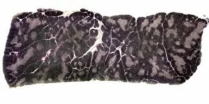
Thymus gland tissue. Light micrograph of a transverse section through tissue from the thymus gland, part of the lymphoid system

Background imageCortex Collection: Cat lymph gland, light micrograph

Cat lymph gland, light micrograph

Background imageCortex Collection: Lymph gland, light micrograph

Lymph gland, light micrograph

Background imageCortex Collection: Inflamed lymph gland, light micrograph

Inflamed lymph gland, light micrograph

Background imageCortex Collection: Purkinje neurons, light micrograph

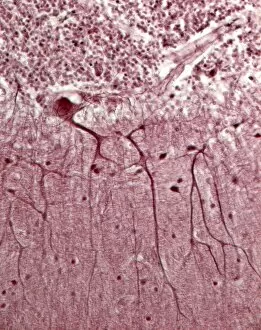
Purkinje neurons, light micrograph


All Professionally Made to Order for Quick Shipping

The cortex, a fascinating part of the brain, is responsible for numerous functions that are essential to our daily lives. One remarkable aspect of the the motor homunculus, a representation of how different parts of our body are controlled by specific areas in the brain. This intricate map showcases the complexity and precision with which movements are coordinated. Purkinje nerve cells in the cerebellum play a crucial role in maintaining balance and coordination. These unique cells have an elaborate branching structure that allows them to transmit information efficiently within this important region of the brain. Examining a vascular bundle under scanning electron microscopy reveals its intricate network, highlighting how blood vessels supply vital nutrients and oxygen to sustain healthy brain tissue. Artwork depicting basal ganglia captures their significance as they contribute to various functions such as movement regulation and decision-making processes. Their interconnectedness emphasizes their influence on overall brain function. Understanding the complex system that ensures proper blood supply to brain tissue is crucial. The delicate balance between oxygen delivery and demand plays a critical role in maintaining optimal cognitive function. A light micrograph showcasing a lime tree stem provides insight into plant anatomy but also serves as a reminder of nature's intricate design parallels with human biology. Similar Purkinje nerve cells can be found within the cerebellum structure when observed under light microscopy. Their distinct appearance further emphasizes their importance in coordinating movements smoothly and precisely. Phantom pain after amputation remains an enigma; artwork capturing this phenomenon highlights its impact on individuals who experience sensations from missing limbs—an intriguing area where neuroscience continues to seek answers. Exploring water fern rhizome under light microscopy offers glimpses into its internal structures, reminding us that biological intricacies extend beyond humans alone—a testament to nature's diversity and complexity. A pine tree stem captured through light microscopy unveils its cellular composition—underscoring similarities between plant tissues and those found within our own bodies—a reflection of shared evolutionary history across species boundaries.